Ultramicrotomes & Cryo-Ultramicrotomes

Whether tissue sample, polymer, rubber, metals or nanoparticles, Leica ultramicrotomes provide extremely thin sections and perfect surface quality in a wide range of applications. From materials science to cancer research, our ultramicrotomes are used for many different kinds of research and quality control all over the world.

Thin sections and perfect surface quality!

Need Assistance?

Contact our team of experts to help you simplify your EM sample preparation.

New UC Enuity Ultramicrotome

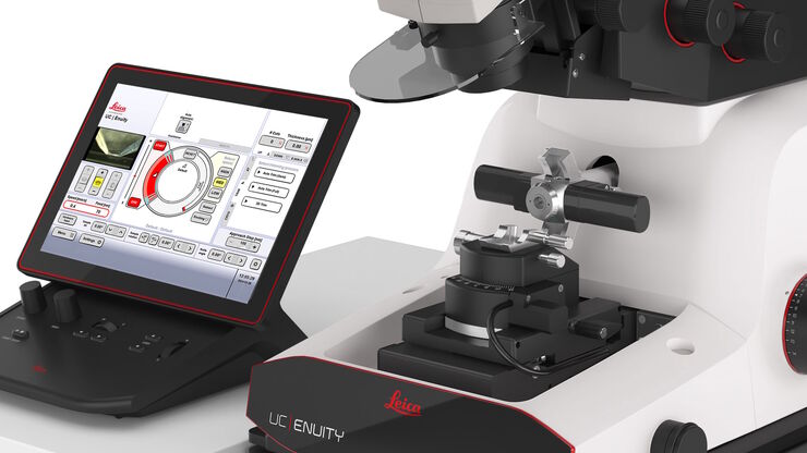
Introducing next generation ultramicrotome that saves you time and resources through its automated setup functions, providing state-of-the-art sectioning quality.

UC Enuity

Follow us on Instagram

No-compromise ergonomics

Users often operate an ultramicrotome for a long period of time. Therefore, fatigue-free operation is a must for both right- and left- handed users. Ergonomic arm rests and generous adjustment options make working with ultramicrotomes from Leica Microsystems more comfortable.

Nanometer precision

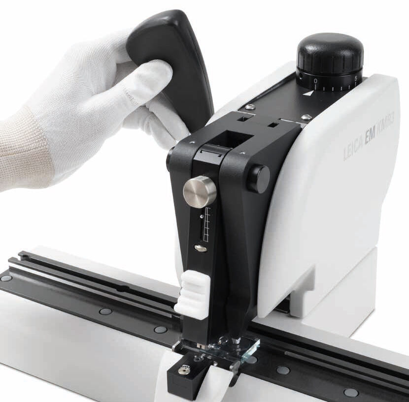
Leica ultramicrotomes guarantee precision and comfort. Thanks to the fully motorized knife stages and a wealth of technical features, even beginners can prepare perfect sections. Make perfect glass knives for perfect ultrathin sections with the EM KMR3 within minutes.

The instruments produce section thicknesses between 10 nm up to 15 µm. Discover the precision mechanics of Leica ultramicrotomes and enjoy highest quality specimen preparation for LM, TEM, SEM, or AFM examination.

ultramicrotomy related articles

Read our latest articles about Ultramicrotomy

The knowledge portal of Leica Microsystems offers scientific research and teaching material on the subjects of microscopy. The content is designed to support beginners, experienced practitioners and scientists alike in their everyday work and experiments.

MORE ARTICLES

Block-face created by automatic trimming under fluorescence. Mammalian cells of interest, stained with CellTrackerTM Green are visualized within the block-face using the UC Enuity equipped with the stereo microscope M205 FA. In the background a carbon finder grid in black is visible. All samples in the article are created by Felix Gaedke, PhD, CECAD, Cologne, Germany.

How to Automatically Obtain Fluorescent Cells of Interest in a Block-face

Block-face created by automatic trimming under fluorescence. Mammalian cells of interest, stained with CellTrackerTM Green are visualized within the block-face using the UC Enuity equipped with the…
UC Enuity

Improve Your Ultramicrotomy Workflow with Automated Sectioning

Discover advanced digital ultramicrotomy tools for fast and accurate automated sectioning. Learn about autoalignment, and efficient sample trimming leveraging 3D µCT data. See application examples…
Camera image during auto alignment. The feedback lines indicate if the correct edges in the image are detected. Green: Vertical center line; Magenta: Upper edge of the light gap; White: Lower edge of the light gap (not visible here, falling together with red line); Red: Knife edge; Blue: Left and right edge of the block face being automatically detected.

Automatic Alignment of Sample and Knife for High Sectioning Quality

Automatic alignment of sample and knife on the ultramicrotome UC Enuity, enabling even untrained users to create ultrathin sections with reduced risk of losing precious sections.
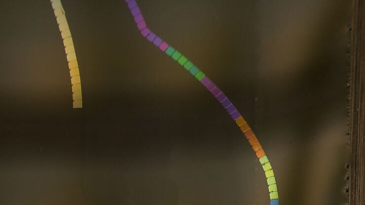
Section ribbons with increasing section thickness - silver to purple ending in blue sections.

High Quality Sectioning in Ultramicrotomy

Discover the significance of achieving high-quality uniform sections with ultramicrotomy for precise imaging in electron microscopy.
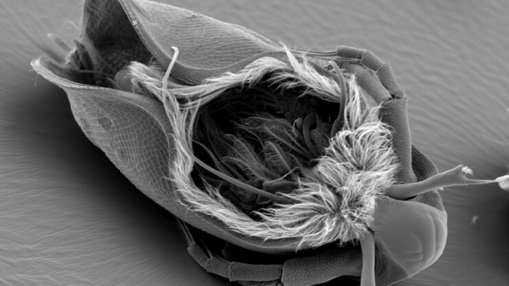
Water Flea Daphnia imaged by Electron Microscopy. Courtesy of Mag. Dr. Gruber, University of Vienna, Austria

Streamline your EM Sample Preparation Workflow for Biological Applications

Master EM sample preparation, including ultramicrotomy, for life sciences in this expert eBook!
Acrylonitrile butadiene styrene (ABS) stained with osmium tetroxide (OsO4), sectioned with a DIATOME diamond knife at room temperature, and then imaged with HAADF TEM.

Ultramicrotomy Techniques for Materials Sectioning

Learn about ultramicrotomy for materials sectioning when investigating polymers and brittle materials with transmission (TEM) or scanning electron microscopy (SEM) or atomic force microscopy.
Ultramicrotomy; ARTOS 3D

Introduction to Ultramicrotomy

When studying samples, to visualize their fine structure with nanometer scale resolution, most often electron microscopy is used. There are 2 types: scanning electron microscopy (SEM) which images the…

Brief Introduction to Specimen Trimming

Before ultrathin sectioning a sample with an ultramicrotome it has to be pre-prepared. For this pre-preparation, special attention must be paid to the sample size (size of the section), location of…
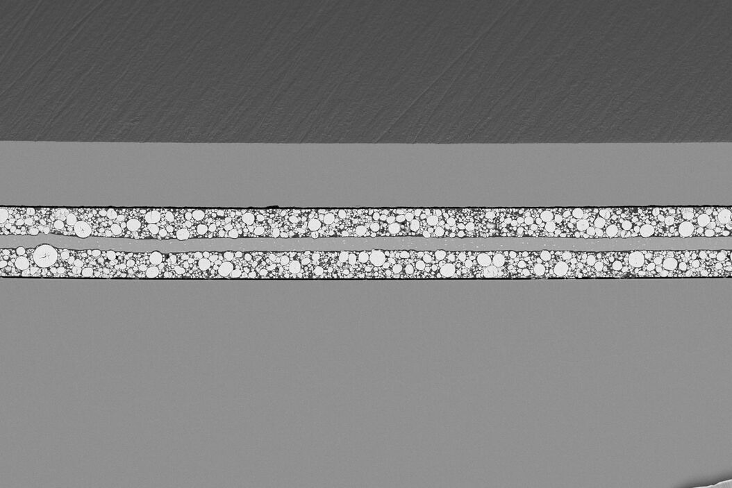
SEM image of the full Li-NMC electrode sample, showing the two porous layers and the metal film at the center of the structure.

Ion Beam Milling Systems

When material specimen surfaces are prepared for SEM or incident light microscopy, the specimen usually undergoes multiple processes until the layer or surface to be analyzed is machined with precision. Leica Microsystems’ workflow solutions for solid state technology cover all steps required for demanding high-quality sample preparation.

Read More

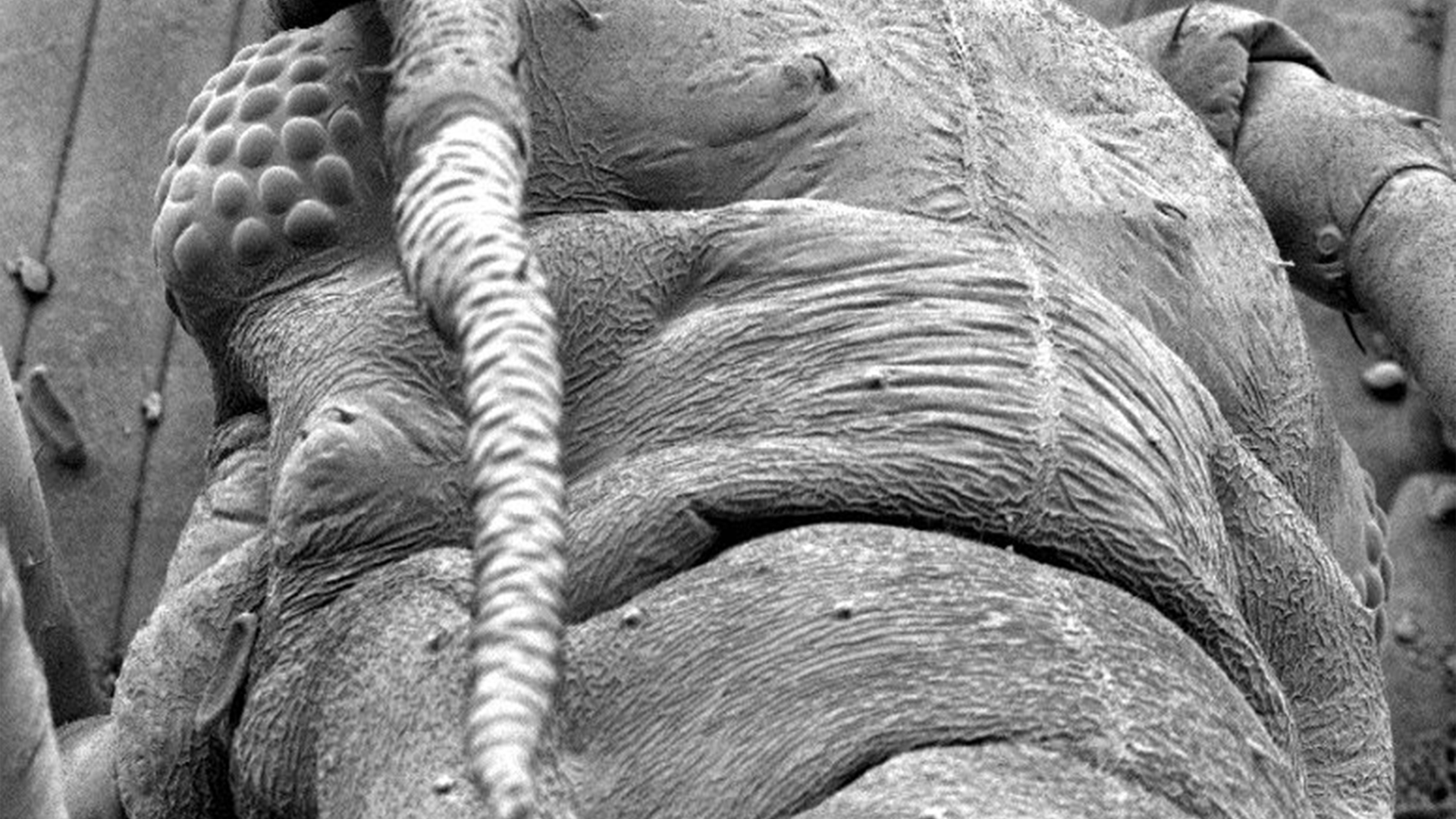
Plant-louse on a wheat leave

Sputter Coater & Freeze Fracture Solutions

To obtain high-quality images of samples with scanning (SEM) or transmission electron microscopy (TEM), your samples need to be conductive to avoid charging. If a sample does not have a high enough conductivity, then you can quickly cover it with a conductive layer using the method of sputter coating. Also, a carbon or e-beam evaporator coating can be used. Such coatings protect the sample, allow enhancing of the EM image contrast, or can act as a TEM-grid support film for small scale samples.

Read More

Cryo Preparation Systems

High pressure freezing is often the preferred method for preserving aqueous samples in their close-to-native state, as it captures the intricate changes in fine structure or cellular dynamics. Leica Microsystems combines high pressure freezing with light stimulation or electrical stimulation: It enables you to visualize highly dynamic processes or the structural changes of samples at a nanometer resolution and with millisecond precision.

Read More

SEM image of the full Li-NMC electrode sample, showing the two porous layers and the metal film at the center of the structure.
Plant-louse on a wheat leave
Scroll to top