Dissecting Microscopes

Dissecting microscopes help you have a better view of a specimen or sample when performing a dissection. You can spend many hours looking through the eyepieces of a dissecting microscope whenever dissections must be done. Leica Microsystems lets you select from a diverse array of microscopes and comprehensive range of dissecting microscope parts and accessories so that you can find the microscope solution that best fits your needs.


Need Assistance?

Contact us for expert advice on the right dissecting microscope for your needs and budget.

A key instrument for your laboratory or classroom

The selection of the right dissecting microscope for the hours you, your lab members, or your students may spend doing dissection is key to long-lasting, satisfactory use.

What should you look for in terms of dissecting microscope magnification and its other properties?

Whether your priority for a dissecting microscope is working distance, magnification, field of view, illumination, high resolution, depth of field, or optical quality, Leica Microsystem can provide the solution that works best for you.

Here are some key factors to consider when choosing a dissecting microscope:

  • Clear view of the specimen’s or sample’s targeted area is important which is best achieved by bright illumination in the microscope’s field of view from proper light sources and high transmission optics;
  • Sufficiently large area of the specimen or sample must be seen with the microscope which is best attained with a large field of view using eyepieces or a camera;
  • Plenty of space to work with utensils under the microscope objective is necessary and this requires a large working distance, the distance between the objective’s front lens and the top of the sample when it is in focus, it normally decreases as the magnification increases;
  • No objects should obstruct your hands and utensils under the microscope and a comfortable head and sitting position while working are important to avoid strain on the neck, back, and arms, so it is useful to have an ergonomic setup;
  • Versatile and easy-to-set-up microscope configuration saves users time, reduces stress, and allows it to be used for multiple tasks.

Do you need to only observe the specimen/sample during dissection or also document your work?

If you need to document the work you do using the dissecting microscope, then integration with a Leica digital camera allows for practical documentation of results.

How many different people need to use the dissecting microscope? How many hours will they work at the microscope?

When it comes to using the dissecting microscope for many hours, it is important to consider ergonomic accessories as they can prevent strain injuries. Depending on the number of different users, it is advisable to have a microscope which can be adjusted to the preference of each user.

Is a dissecting microscope the same thing as a microdissection microscope?

No, normally microdissection is performed with a laser. The microscope which does this is known as a laser microdissection (LMD) microscope. It enables the isolation of specific areas of tissue, single cells, or even subcellular structures of a specimen.

Stroke and Cancer Research

Improvement in stroke and cancer treatment methods depend on advances made from laboratory research. Rats and mice are the most abundant mammalian model organisms used for such research. An indispensable tool for any stroke or cancer laboratory is a dissecting microscope.

Read More

Education

Leica dissecting microscopes and dissecting microscope parts are used for education in classrooms and laboratories of schools and universities around the world. The microscopes offer high-quality optics and illumination and sufficient space under the objective for easy access to the specimen or sample. They are also student-friendly in terms of setup and use.

Read More

Frequently Asked Questions Dissecting Microscopes

Show answer What is a dissecting microscope and what is it used for?

A dissecting microscope, also known as a stereo microscope, is used to perform dissection of a specimen or sample. It simply gives the person doing the dissection a magnified, 3-dimensional view of the specimen or sample so more fine details can be visualized.

Show answer Why is a stereo microscope also called a dissecting microscope?

Stereo microscopes are always used to perform dissections of a specimen or sample, therefore, they can also be called dissecting microscopes. As stereo microscopes use 2 light channels, 1 for each eye, they give the observer a magnified, 3-dimensional view of the specimen or sample. This ability makes them practical to use for dissection.

Show answer Other than Leica cameras, can I mount 3rd party SLR/DSLR cameras to a Leica microscope or macroscope for that matter?

We have adapters for all C-mount compatible cameras.

Show answer Are Leica microscopes only compatible with the Leica Application Suite software? Is integration with a 3rd party software possible?

Yes, use of a 3rd party software is possible: https://www.splashtop.com/classroom

Show answer What type of accessories does my dissecting microscope come with? What will I need to purchase separately?

Leica microscopes are modular and shipped in the configuration that best fits your stated needs. In case your needs change later, you can always upgrade your microscope by adding available accessories.

Show answer What kinds of modular accessories are available to extend the range of use of my dissecting microscope?

There are a lot of microscope accessories. Please get in contact with your local Leica sales representative or visit: https://www.leica-microsystems.com/products/microscope-accessories

Show answer How are ergonomic accessories designed to reduce repetitive strain injuries during long working hours?

Ergonomic accessories help users perform dissections in comfort, even if they spend long hours working with a microscope. Leica accessories allow you to adapt the dissecting microscope to your individual needs, allowing you to dissect more efficiently. There are a lot of ergonomic accessories for Leica microscopes. Please contact your local Leica sales representative for more details or visit: https://www.leica-microsystems.com/products/microscope-accessories/ergonomics

Related Articles

Read our latest articles about Dissecting Microscopes

The knowledge portal of Leica Microsystems offers scientific research and teaching material on the subjects of microscopy. The content is designed to support beginners, experienced practitioners and scientists alike in their everyday work and experiments.

MORE ARTICLES

Automated Laser Microdissection for Proteome Analysis

Deep Visual Proteomics Provides Precise Spatial Proteomic Information

Despite the availability of imaging methods and mass spectroscopy for spatial proteomics, a key challenge that remains is correlating images with single-cell resolution to protein-abundance…
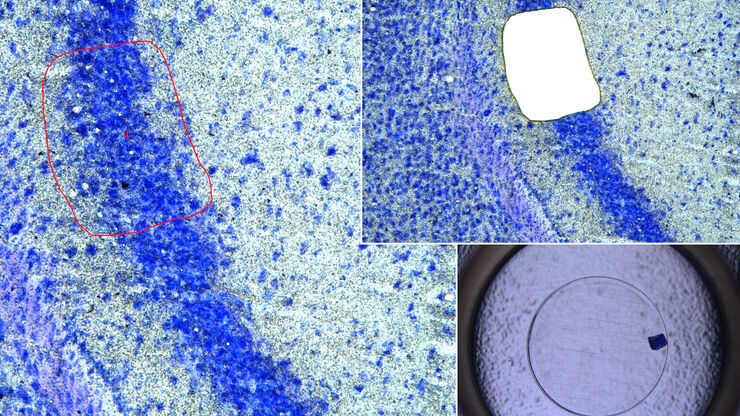
Image of a 12-μm thick brain section, which was stained with Toluidin blue, before dissection. It was taken with a microscope using a 63x objective.

An Introduction to Laser Microdissection

The heterogeneity of histological and biological specimens often requires isolation of specific single cells or cell groups from surrounding tissue before molecular biology analysis can be carried…
Mouse brain (left) microdissected with a 10x objective (upper right). Inspection of the collection device (lower right).

Molecular Biology Analysis facilitated with Laser Microdissection (LMD)

Extracting biomolecules, proteins, nucleic acids, lipids, and chromosomes, as well as extracting and manipulating cells and tissues with laser microdissection (LMD) enables insights to be gained into…
Brightfield image of a pig liver stained with hematoxylin-eosin (HE).

Spatial Metabolomics: Exploring Tumor Complexity and Therapeutic Insights

In cancer research, it is vital to understand the interaction between tumor cells and their microenvironment, as the tumor microenvironment influences tumor progression significantly. Spatial…
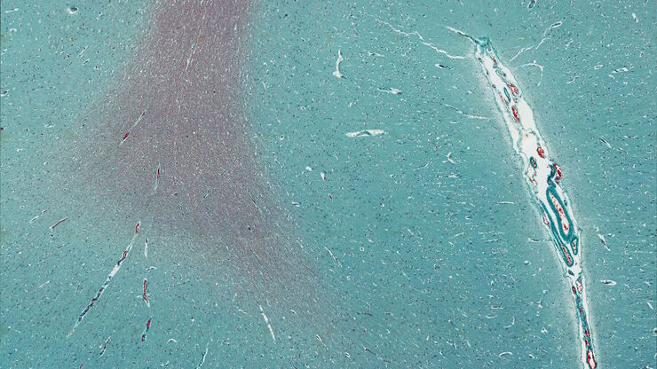
Mosaic scan of a Masson-Goldner stained cat brain. Magnification: 20x.

Lipidomics Analysis of Sparse Cells based on Laser Microdissection

Delve into cellular intricacies with high-coverage targeted lipidomics analysis of sparse cells. This advanced method, integrating Laser Microdissection (LMD) and Liquid Chromatography-Mass…
Image of murine dopaminergic neurons which have been marked for laser microdissection (LMD).

Neuron Isolation in Spatial Context with Laser Microdissection (LMD)

After Alzheimer’s disease, Parkinson’s is the second most common progressive neurodegenerative disease. Before the first symptoms manifest, up to 70% of dopamine-releasing neurons in the mid-brain…

How did Laser Microdissection enable Pioneering Neuroscience Research?

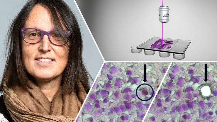
Dr. Marta Paterlini, a Senior Scientist at the Karolinska Institute, shares her experience of using laser microdissection (LMD) in groundbreaking research into adult human neurogenesis and offers…

Laser Microdissection Protocols for Tissue and Cell Isolation - Download free eBook

In this Bio-protocol Selections, we present a collection of open-access, detailed methods papers using LCM to purify and isolate tissues and cells from plants, mouse embryos, cancer cells, neurons,…
Single cell datasets

Exploring Subcellular Spatial Phenotypes with SPARCS

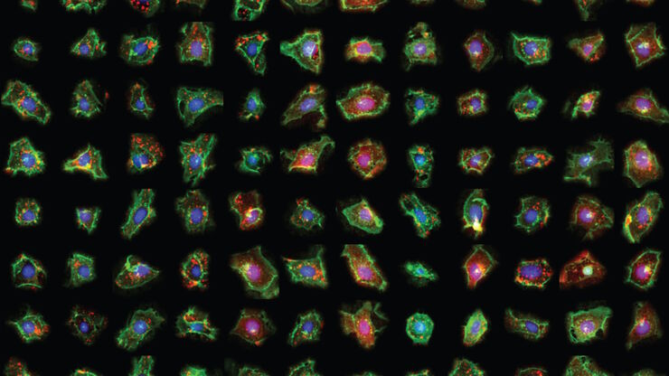
Discover spatially resolved CRISPR screening (SPARCS), a platform for microscopy-based genetic screening for spatial subcellular phenotypes at the human genome scale.
How is microscopy used in spatial biology - Teaserimage

How is Microscopy Used in Spatial Biology? A Microscopy Guide

Different spatial biology methods in microscopy, such as multiplex imaging, are helping to better understand tissue landscapes. Learn more in this microscopy guide.
Scroll to top