GLOW800
Surgical Microscopes
Products
Home
Leica Microsystems
GLOW800 Augmented Reality Fluorescence Application
Vascular Surgery. Augmented
Read our latest articles
Aneurysm Clipping: Assessing Perforators in Real-Time with AR Fluorescence
This article covers two aneurysm clipping cases highlighting the clinical benefits of GLOW800 Augmented Reality Fluorescence application in neurosurgery, based on insights from Prof. Tohru Mizutani,…
Augmented Reality: Transforming Neurosurgical Procedures
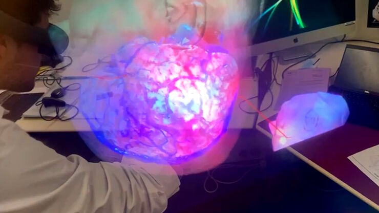
In this ebook, you will explore the exciting advances that Augmented Reality (AR) brings to the field of neurosurgery. This comprehensive guide, including explanatory videos, addresses key questions…
Enhancing Neurosurgery Teaching
Learn about the Serious Game in Intraoperative Neurosurgery and how it supports neurosurgical teaching and the acquisition of decision-making skills.
Use of AR Fluorescence in Neurovascular Surgery
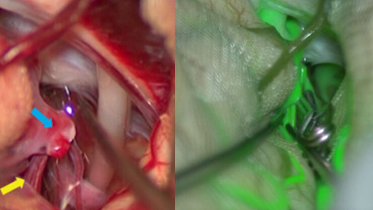
Learn about the use of GLOW800 Augmented Reality in neurovascular surgery through clinical cases and videos, including aneurysm and tumor resection cases.
3D, AR & VR for Teaching in Neurosurgery
Discover the evolution of neurosurgical teaching and how 3D, Augmented Reality and Virtual Reality can help better learn anatomy and acquire surgical skills.
AR Fluorescence in Aneurysm Clipping and AVM Surgery
Discover how GLOW800 Augmented Reality fluorescence supports neurovascular surgical procedures and in particular aneurysm clipping and AVM surgery.
Digitalization in Neurosurgical Planning and Procedures
Learn about Augmented Reality, Virtual Reality and Mixed Reality in neurosurgery and how they can help overcome challenges.
Augmented Reality Assisted Navigation in Neuro-Oncological Surgery
In neuro-oncological surgery, new technologies such as Augmented Reality are helping to improve surgical precision enabling a precise trajectory, conformational resection, the absence of collateral…
How AR Helps in the Surgical Treatment of Moyamoya Disease
Moyamoya disease is a rare chronic occlusive cerebrovascular disorder characterized by progressive stenosis in the terminal portion of the internal carotid artery and an abnormal vascular network at…
Using GLOW800 AR in Radial Forearm Flap Free Phalloplasty
In this video, Chief Microsurgeon Professor Küntscher and his team perform a radial forearm free flap phalloplasty and use ICG fluorescence imaging to show the blood flow in the whole flap from the…
How AR Surgery Benefits Radial Forearm Free Flap Phalloplasty
The goal of penile reconstruction is to provide an aesthetic penoid with tactile and erogenous sensation, so the patient can have sexual intercourse and void standing.1 Currently, the radial forearm…
Free Flap Procedures in Oncological Reconstructive Surgery
Free flap surgery is considered the gold standard for breast, head and neck reconstructions for cancer patients. These procedures, which enable functional and aesthetic rehabilitation, can be quite…
Advances in Oncological Reconstructive Surgery
Decision making and patient care in oncological reconstructive surgery have considerably evolved in recent years. New surgical assistance technologies are helping surgeons push the boundaries of what…
How Augmented Reality is Transforming Vascular Neurosurgery
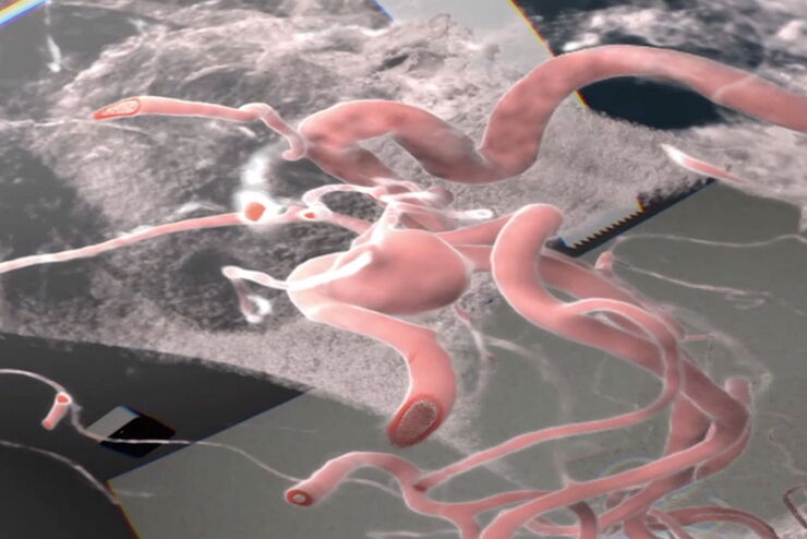
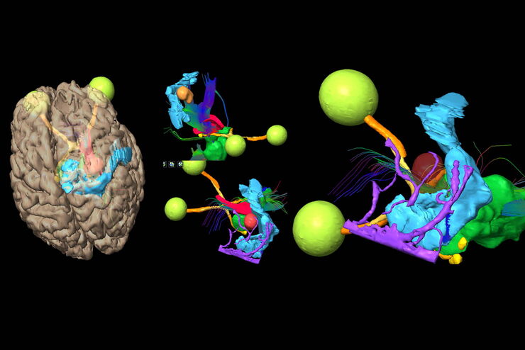
Augmented Reality is changing surgery, with new information helping to improve the precision and safety of procedures. This is especially true in vascular neurosurgery where Augmented Reality is…
GLOW800 Augmented Reality Fluorescence in AVM (Arteriovenous Malformation) Treatment
In this case study Prof. Dr. Feres Chaddad talks about the treatment of AVMs. It illustrates how the Augmented Reality Fluorescence GLOW800 can help surgeons during microsurgical resection by…
Neurovascular Surgery & Augmented Reality Fluorescence
Vascular neurosurgery is highly complex. Surgeons need to be able to rely on robust anatomical information. As such, visualization technologies play an essential role.
Prof. Nils Ole Schmidt is a…
Oncological Reconstructive Surgery with the Leica M530 OHX Microscope
Precision is essential in oncological reconstructive surgery, in particular when it relies on free flap techniques. Microsurgical microscopes provide optimal visualization and help streamline the…
Oncological Reconstructive Surgery: Why Use a Microscope
Recent advances in microsurgery are enhancing breast reconstruction for oncology patients, allowing both functional and aesthetic rehabilitation. More and more surgeons are adopting surgical…
Neurosurgery with Heads-up Display
In the following video interviews Prof. Dr. Raphael Guzman, Vice Chairman of the Department of Neurosurgery at the University Hospital in Basel, Switzerland, talks about his experience in heads-up…
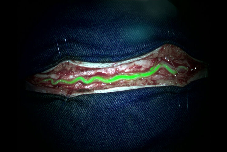
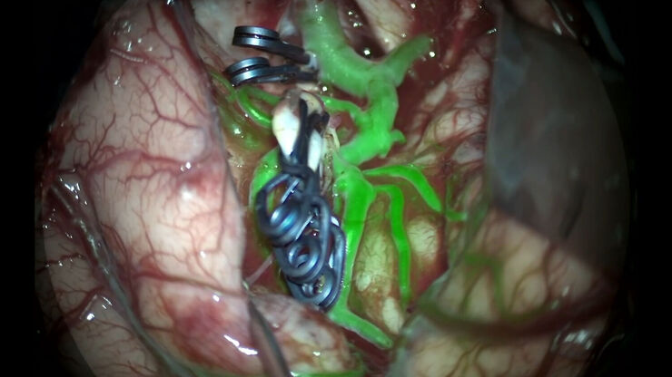
Augmented Reality (AR) Fluorescence Image Gallery
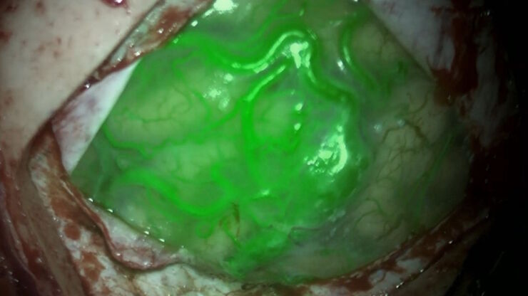
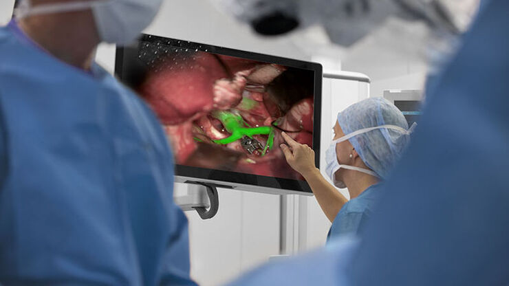
Building on a decade of leadership in fluorescence imaging technology, GLOW800 AR fluorescence is the first of many modalities based on the proprietary GLOW AR platform.
The sophisticated imaging…
Clinical Uses in Cerebrovascular and Skull Base Neurosurgery
In this webinar Dr. Bendok and Dr. Morcos explain how Augmented Reality and Fluorescence can enhance visualization and support surgical decision making. They present first-hand experience of the GLOW…
Surgical Microscopes: Key Factors for OR Nurses
Operating room (OR) nurses are vital to the surgery process. An OR Nurse Manager explains the key surgical microscope features to facilitate their work.
GLOW800 Augmented Reality Fluorescence in Aneurysm Treatment
This case study from Prof. Dr. Feres Chaddad talks about the treatment of unruptured MCA (middle cerebral artery) and PCOM (posterior communicating artery) aneurysms with microsurgical clipping. It…
The Future of Fluorescence in Vascular Neurosurgery
In vascular neurosurgery, surgical microscopes are used to provide a magnified and illuminated view of the surgical field. Although surgeons benefit greatly from the superb image quality and optical…
Fields of Application
Neurosurgery
Neurosurgery microscopes from Leica Microsystems provide high-quality optics and engineering for cranial, spine, multi-disciplinary and other microsurgery disciplines.
Plastic & Reconstructive Surgery
Whether you need a premium surgical microscope or a cost-effective option for multidisciplinary use, Leica Microsystems offers customizable options for a wide range of microsurgical applications.
