
Content
Polarized light microscopy is used for a wide range of applications in the earth sciences and quality control in various industries. A variety of polarizing microscope images from geological and industrial applications are shown in this gallery.
- Earth Sciences: geology, petrography, mineralogy, crystal structure characterization, asbestos analysis, and coal analysis (vitrinite reflectance)
- Quality control: glass (stress birefringence or inclusions), plastics and polymers (stress birefringence), textiles and fibers, electronic displays, and examination of liquid crystals.
Asbestos
The term asbestos covers 6 natural silicate minerals with different properties. Each one consists of long, flexible crystal fibers. Asbestos was used as a building material many years ago before it was known to be a health hazard. If found in an old building, depending on the type, costly disposal may be necessary.

Right: Same actinolite sample imaged with crossed polarizers. The actinolite fibers show prominent colors from birefringence which clearly distinguish them from glass fibers (no birefringence).
Images recorded with a DM4 P microscope using transmitted light, 20x Plan Fluotar objective, and polarizers.

Image recorded with a DM4 P microscope using transmitted light, 20x Plan Fluotar objective, and polarizers.

Images recorded with a DM4 P microscope using transmitted light, 20x N Plan DS (dispersion staining) objective, and polarizers.

Image recorded with a DM4 P microscope using transmitted light, 20x N Plan DS (dispersion staining) objective, and polarizers.
Ore
Ores are natural rocks that contain valuable minerals. Ores are typically gathered by mining and then further processed so the desired minerals are extracted. Metal ores are often oxides, sulfides, or silicates. The crystal structure and composition of ores can be characterized with polarized light microscopy.

Image recorded with a DM4 P microscope using transmitted light, 40x Plan Fluotar objective, and crossed polarizers.
Fibers
Polarization microscopy quickly provides useful information about fibers, such as human hair or natural (e.g., wool, cotton, silk, etc.) and synthetic (e.g., polymers like polyamide nylon, polyester, rayon, etc.) fibers. Often this information can help distinguish them.

Right: Same nylon fibers imaged with crossed polarizers show typical higher order birefringence colors.
Images recorded with a DM4 P microscope using transmitted light, 20x Plan Fluotar objective, and polarizers.

Right: Same Perlon®* fiber imaged with crossed polarizers showing high order birefringence colors.
Images recorded with a DM4 P microscope using transmitted light, 20x Plan Fluotar objective, and polarizers.
* All product and company names are trademarks™ or registered® trademarks of their respective holders. Use of them does not imply any affiliation with or endorsement by them.

Right: Same wool fiber imaged with crossed polarizers showing no clear birefringence colors.
Images recorded with a DM4 P microscope using transmitted light, 20x Plan Fluotar objective, and polarizers.
Geology
The crystal structure and composition of rocks and minerals can be characterized with polarized light. The examination of rocks with polarized light microscopy normally requires thin sections, approximately 25 µm in thickness, to be produced.

Image recorded with a DM4 P microscope using transmitted light, 5x Plan Fluotar objective, and polarizers.

Right: Same basalt section imaged with crossed polarizers. Large crystals show higher birefringence colors (pyroxene & olivine). Strip-shaped plagioclase are clearly visible due to their shape and low birefringence.
Images recorded with a DM4 P microscope using transmitted light, 10x Plan Fluotar objective, and polarizers.

Image recorded with a DM4 P microscope using transmitted light, 40x Plan Fluotar objective, and polarizers.

Right: Same symplectic structure in eclogite imaged with crossed polarizers.
Images recorded with a DM4 P microscope using transmitted light, 20x Plan Fluotar objective, and polarizers.
Coal
In coal petrography, special oil immersion objectives can be used to differentiate between different coal components (macerals). For these incident light investigations, the coal samples are ground to a high polish. The examination of the coal components allows conclusions to be drawn about the history of the coal deposit, energy content of the coal, etc. The differentiation of macerals is done essentially according to their shape, reflection strength (brightness), and fluorescence characteristics.

Image recorded with a DM4 P microscope using 20x Oil N Plan objective and XLR polarizers.

Image recorded with a DM4 P microscope using 50x Oil N Plan objective and XLR polarizers.
Conoscopy
An optical technique for observing transparent samples with light that converges into a cone. All directions in which the light propagates can be observed at the same time. Conoscopy is performed with a polarized light microscope using a Bertrand lens. Conoscopy is useful for evaluating the optical properties, for example the number of optical axes, of anisotropic materials.

Right: Conoscopic image of the same calcite sample with linear polarized light. The calcite section is perpendicular to the optical axis.
Images recorded with a DM4 P microscope using transmitted light, conoscopy, 63x N Plan objective, and polarizers.

Right: Conoscopic image of the same muscovite sample with circularly polarized light. Position of the optical axes clearly determined with circular polarization.
Images recorded with a DM4 P microscope using transmitted light, conoscopy, 63x N Plan objective, and polarizers.
Plastics
Residual stress or inhomogeneities in plastics/polymers may occur when they are manufactured. Often, the presence of such stress is unseen and can become a problem that leads to failure. Detection of stress or inhomogeneities with quality control during production can help minimize it. Stressed regions and inhomogeneities present in plastics are visible as bands of multiple colors with polarized light microscopy.

Right: Same polypropylene sample imaged with crossed polarizers. Inhomogeneities that may have occurred during the production process are revealed.
Images recorded with a DM4 P microscope using 2.5x Plan Fluotar objective and polarizers.

Right: Same polyethylene film imaged with crossed polarizers. The image shows more distinctly the inhomogeneities.
Images recorded with a DM4 P microscope using 2.5x Plan Fluotar objective and polarizers.

Right: Same polyethylene film imaged with crossed polarizers and a lambda plate. The size and distribution of the spherulites have a decisive influence on the film’s properties.
Images recorded with a DM4 P microscope using 20x Plan Fluotar objective and polarizers.
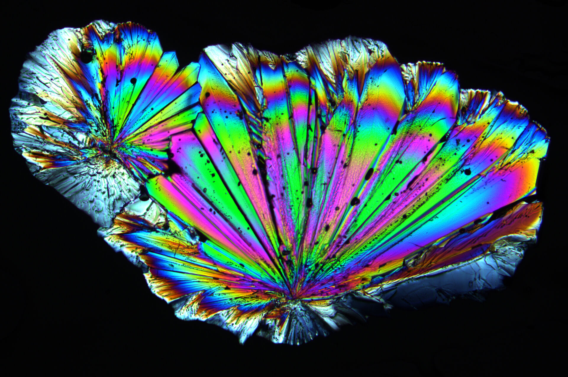
Organic Materials
The crystal structure and composition of organic materials can be characterized with polarized light microscopy.

Image recorded with a DM4 P microscope using transmitted light, 20x Plan Fluotar objective, and polarizers.

Image recorded with a DM4 P microscope using transmitted light, 10x Plan Fluotar objective, and polarizers.

Image recorded with a DM4 P microscope using transmitted light, 20x Plan Fluotar objective, and polarizers.

Image recorded with a DM4 P microscope using transmitted light, 10x Plan Fluotar objective, and polarizers.
 and oblique (right) brightfield illumination using a Leica compound microscope. The defect on the wafer surface is clearly more visible with oblique illumination.")


