Corporate Communications

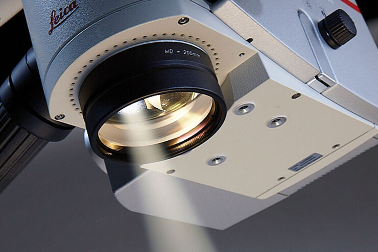
Leica Microsystems develops and manufactures microscopes and scientific instruments for the analysis of microstructures and nanostructures.
We offer scientific research and teaching material on the subjects of microscopy. The content is designed to support beginners, experienced practitioners and scientists alike in their everyday work and experiments. Explore interactive tutorials and application notes, discover the basics of microscopy as well as high-end technologies.
Follow us
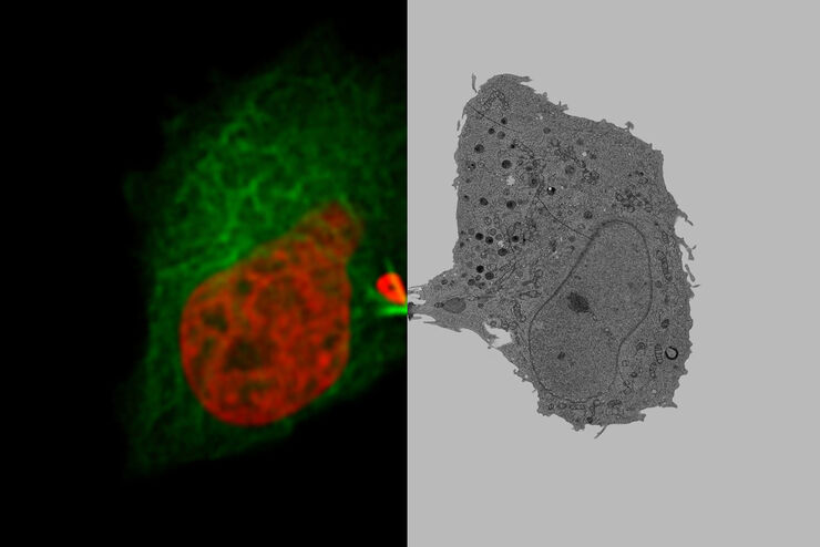
Putting Dynamic Live Cell Data into the Ultrastructural Context
With workflow Coral Life, searching for a needle in the haystack is a thing of the past. Take advantage of correlative light and electron microscopy to identify directly the right cell at the right…
AI in Microscopy Webinar
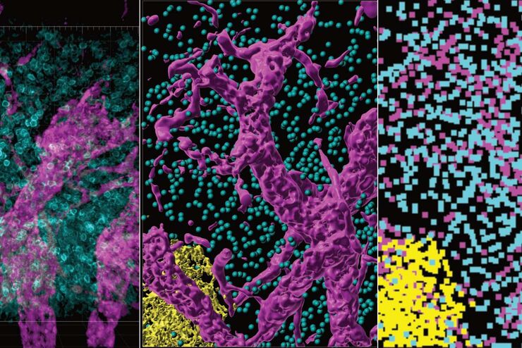
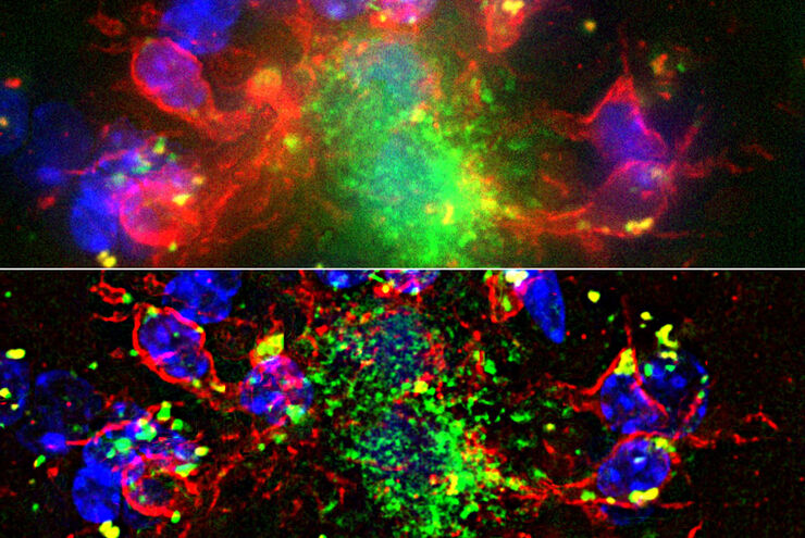
We demonstrate residual channel attention networks for restoring and enhancing volumetric time-lapse (4D) fluorescence microscopy data.
 and astrocytes (green) in a cortical spheroid derived from human induced pluripotent stem cells.")
Download The Guide to Live Cell Imaging
In life science research, live cell imaging is an indispensable tool to visualize cells in a state as in vivo as possible. This E-book reviews a wide range of important considerations to take to…

Regulators of Actin Cytoskeletal Regulation and Cell Migration in Human NK Cells
Dr. Mace will describe new advances in our understanding of the regulation of human NK cell actin cytoskeletal remodeling in cell migration and immune synapse formation derived from confocal and…

Adding Dimensions to Multiplex Molecular Imaging
Molecular imaging of living specimens offers a means to draw upon the growing body of high-throughput molecular data to better understand the underlying cellular and molecular mechanisms of complex…
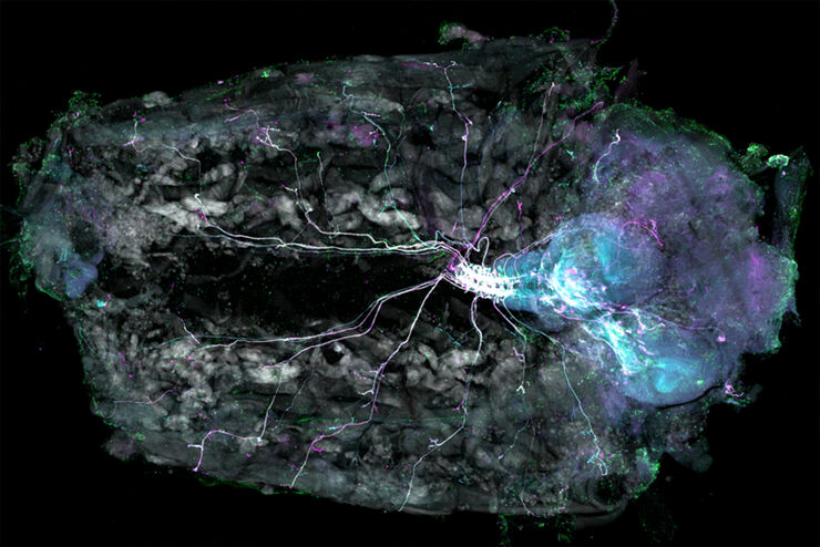
Understanding Motor Sequence Generation Across Spatiotemporal Scales
We have developed a microscopy-based pipeline to characterize a developmentally critical behavior at the pupal stage of development, called the ecdysis sequence. We study brain-wide neuronal activity…

Benefits of TauContrast to Image Complex Samples
In this interview, Dr. Timo Zimmermann talks about his experience with the application of TauSense tools and their potential for the investigation of demanding samples such as thick samples or…

Fast, High-quality Vitrification with the EM ICE High Pressure Freezer
The EM ICE High Pressure Freezer was developed with a unique freezing principle and uses only a single pressurization and cooling liquid: liquified nitrogen (LN2). This design enables three major…

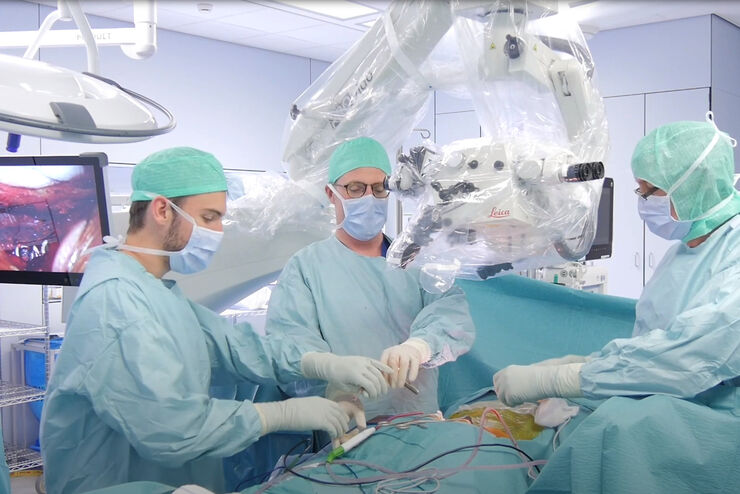
Advanced Visualization in ENT Surgery
ENT surgery poses unique challenges. It requires excellent visualization, both in terms of magnification and illumination. As such, choosing the right operating microscope is critical.
Advanced Visualization in Spine Surgery
There have been meaningful advances in spine surgery in recent years. Minimally Invasive Spine Surgery (MISS) techniques provide significant benefits, helping to minimize surgical trauma, improve…
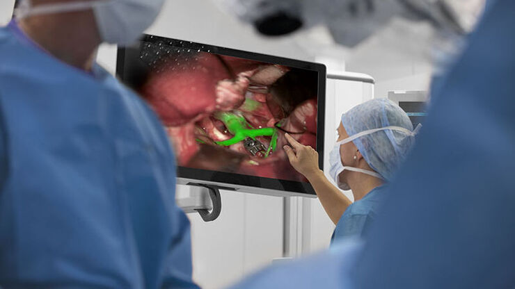
How Augmented Reality is Transforming Vascular Neurosurgery
Augmented Reality is changing surgery, with new information helping to improve the precision and safety of procedures. This is especially true in vascular neurosurgery where Augmented Reality is…
Overcoming Ophthalmologic Surgery Challenges
Ophthalmology surgical procedures involving both the anterior and posterior segment can be particularly challenging. Good visualization is required to operate with precision and confidence.
Prof.…

Advanced Visualization in Maxillofacial Plastic and Reconstructive Surgery
Plastic and Reconstructive Surgery is very demanding. Surgical microscopes play an important role, helping to ensure flaps are well vascularized.
Dr. Christine Bach is a Plastic and Reconstructive…
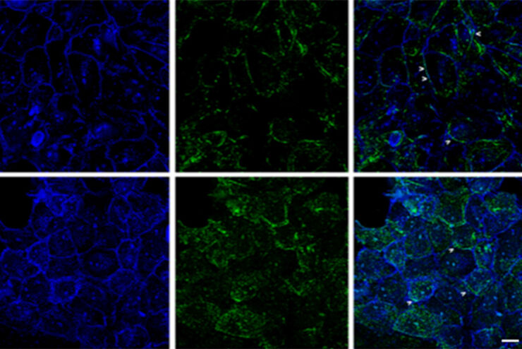
Improvement of Imaging Techniques to Understand Organelle Membrane Cell Dynamics
Understanding cell functions in normal and tumorous tissue is a key factor in advancing research of potential treatment strategies and understanding why some treatments might fail. Single-cell…

Neurovascular Surgery & Augmented Reality Fluorescence
Vascular neurosurgery is highly complex. Surgeons need to be able to rely on robust anatomical information. As such, visualization technologies play an essential role.
Prof. Nils Ole Schmidt is a…

Advancing Cell Biology with Cryo-Correlative Microscopy
Correlative light and electron microscopy (CLEM) advances biological discoveries by merging different microscopes and imaging modalities to study systems in 4D. Combining fluorescence microscopy with…

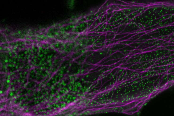
Image Gallery: THUNDER Imager
To help you answer important scientific questions, THUNDER Imagers eliminate the out-of-focus blur that clouds the view of thick samples when using camera-based fluorescence microscopes. They achieve…

Hand-Held OCT Clinical & Research Applications
Hand-held Optical Coherence Tomography (OCT) has revolutionized pediatric ophthalmology and has had a significant impact on ophthalmology in general. The Leica Envisu C2300 hand-held OCT allows to…
From Organs to Tissues to Cells: Analyzing 3D Specimens with Widefield Microscopy
Obtaining high-quality data and images from thick 3D samples is challenging using traditional widefield microscopy because of the contribution of out-of-focus light. In this webinar, Falco Krüger…

Plastic & Reconstructive Surgery: Why Use a Microscope
Plastic and Reconstructive Surgery procedures can be delicate. Visualization solutions play an essential role, allowing to perform the surgery in the best conditions. And more and more plastic…

Minimally Invasive Spine Surgery: Improving Precision and Accuracy with Microscopes
Spine surgery is extremely delicate and requires extensive training and experience. Innovative visualization technologies can also help achieve better outcomes allowing to see more and have a clearer…

A Guide to Model Organisms in Research
A model organism is a species used by researchers to study specific biological processes. They have similar genetic characteristics to humans and are commonly used in research areas such as genetics,…
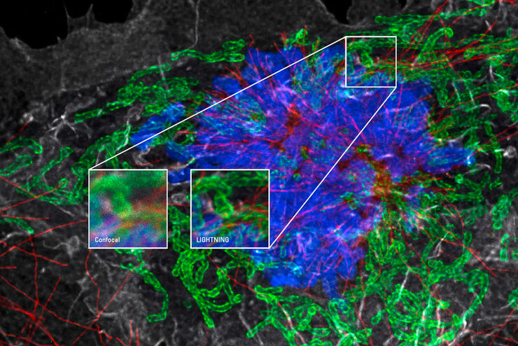
Obtain Maximum Information from your Specimen with LIGHTNING
LIGHTNING is an adaptive process for extraction of information that reveals fine structures and details, otherwise simply not visible, fully automatically. Unlike traditional technologies, that use a…
Advanced Techniques in Cataract and Refractive Surgery
In this webinar Dr. Thompson and Dr. Moshirfar will explain how Leica microscopes aid in procedures such as Centration of Multifocal IOLs and corneal inlays such as Kamra and Lenticular Grafts used in…
Clinical Uses in Cerebrovascular and Skull Base Neurosurgery
In this webinar Dr. Bendok and Dr. Morcos explain how Augmented Reality and Fluorescence can enhance visualization and support surgical decision making. They present first-hand experience of the GLOW…

Surgical Microscopes: Key Factors for OR Nurses
Operating room (OR) nurses are vital to the surgery process. An OR Nurse Manager explains the key surgical microscope features to facilitate their work.
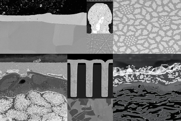
Introduction to Ion Beam Etching with the EM TIC 3X
In this article you can learn how to optimize the preparation quality of your samples by using the ion beam etching method with the EM TIC 3X ion beam milling machine. A short introduction of the…
Computational Clearing - Enhance 3D Specimen Imaging
This webinar is designed to clarify crucial specifications that contribute to THUNDER Imagers' transformative visualization of 3D samples and improvements within a researcher's imaging-related…

STELLARIS White Light Lasers
When it comes to choosing fluorescent probes for your multi-color experiments, you shouldn’t have to compromise. Now you can advance beyond conventional excitation sources that limit your fluorophore…
How to Drape an Overhead Surgical Microscope
The tutorial features the Leica ARveo digital Augmented Reality microscope for complex neurosurgery. The procedure also applies to the Leica M530 OHX, OH6, OH5 and OH4.
