Corporate Communications

Leica Microsystems develops and manufactures microscopes and scientific instruments for the analysis of microstructures and nanostructures.
We offer scientific research and teaching material on the subjects of microscopy. The content is designed to support beginners, experienced practitioners and scientists alike in their everyday work and experiments. Explore interactive tutorials and application notes, discover the basics of microscopy as well as high-end technologies.
Follow us
Advanced Visualization in Maxillofacial Plastic and Reconstructive Surgery
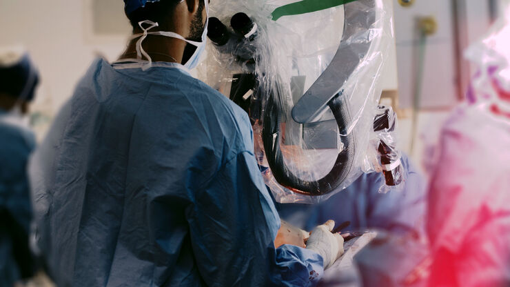
Plastic and Reconstructive Surgery is very demanding. Surgical microscopes play an important role, helping to ensure flaps are well vascularized.
Dr. Christine Bach is a Plastic and Reconstructive…
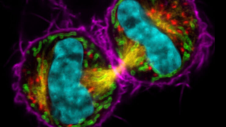
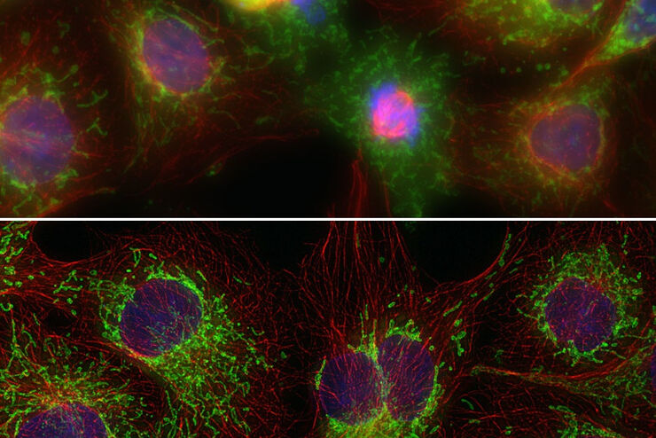
Improvement of Imaging Techniques to Understand Organelle Membrane Cell Dynamics
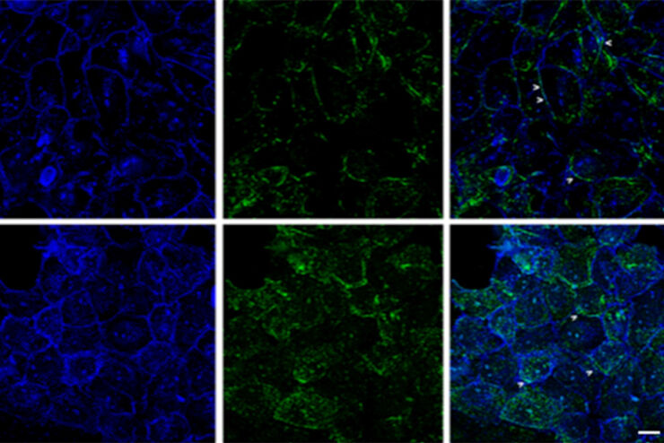
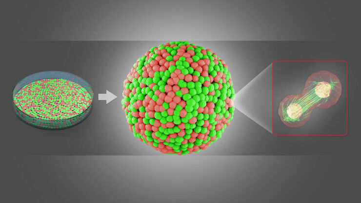
Understanding cell functions in normal and tumorous tissue is a key factor in advancing research of potential treatment strategies and understanding why some treatments might fail. Single-cell…
Neurovascular Surgery & Augmented Reality Fluorescence
Vascular neurosurgery is highly complex. Surgeons need to be able to rely on robust anatomical information. As such, visualization technologies play an essential role.
Prof. Nils Ole Schmidt is a…
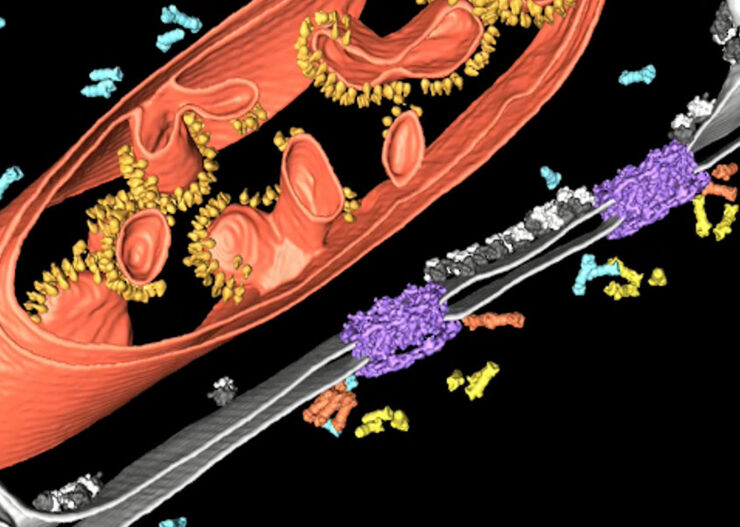
Advancing Cell Biology with Cryo-Correlative Microscopy
Correlative light and electron microscopy (CLEM) advances biological discoveries by merging different microscopes and imaging modalities to study systems in 4D. Combining fluorescence microscopy with…
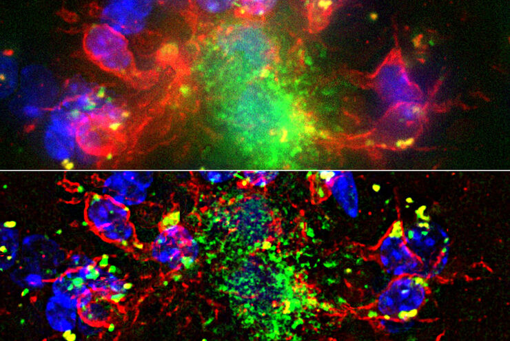
Image Gallery: THUNDER Imager
To help you answer important scientific questions, THUNDER Imagers eliminate the out-of-focus blur that clouds the view of thick samples when using camera-based fluorescence microscopes. They achieve…
Hand-Held OCT Clinical & Research Applications
Hand-held Optical Coherence Tomography (OCT) has revolutionized pediatric ophthalmology and has had a significant impact on ophthalmology in general. The Leica Envisu C2300 hand-held OCT allows to…
From Organs to Tissues to Cells: Analyzing 3D Specimens with Widefield Microscopy
Obtaining high-quality data and images from thick 3D samples is challenging using traditional widefield microscopy because of the contribution of out-of-focus light. In this webinar, Falco Krüger…
Plastic & Reconstructive Surgery: Why Use a Microscope
Plastic and Reconstructive Surgery procedures can be delicate. Visualization solutions play an essential role, allowing to perform the surgery in the best conditions. And more and more plastic…
Minimally Invasive Spine Surgery: Improving Precision and Accuracy with Microscopes
Spine surgery is extremely delicate and requires extensive training and experience. Innovative visualization technologies can also help achieve better outcomes allowing to see more and have a clearer…
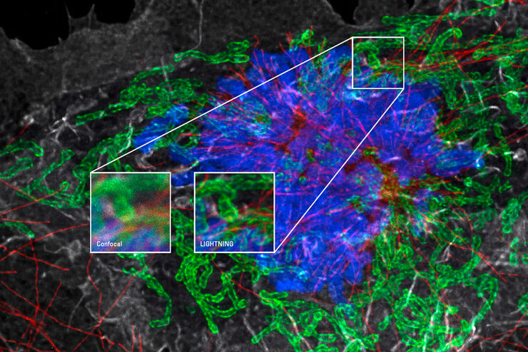
Obtain Maximum Information from your Specimen with LIGHTNING
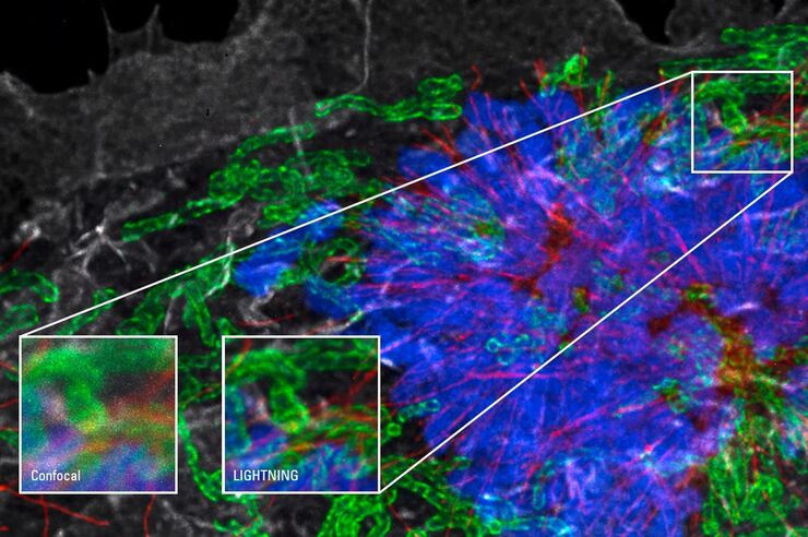
LIGHTNING is an adaptive process for extraction of information that reveals fine structures and details, otherwise simply not visible, fully automatically. Unlike traditional technologies, that use a…
Advanced Techniques in Cataract and Refractive Surgery
In this webinar Dr. Thompson and Dr. Moshirfar will explain how Leica microscopes aid in procedures such as Centration of Multifocal IOLs and corneal inlays such as Kamra and Lenticular Grafts used in…
Clinical Uses in Cerebrovascular and Skull Base Neurosurgery
In this webinar Dr. Bendok and Dr. Morcos explain how Augmented Reality and Fluorescence can enhance visualization and support surgical decision making. They present first-hand experience of the GLOW…
Surgical Microscopes: Key Factors for OR Nurses
Operating room (OR) nurses are vital to the surgery process. An OR Nurse Manager explains the key surgical microscope features to facilitate their work.
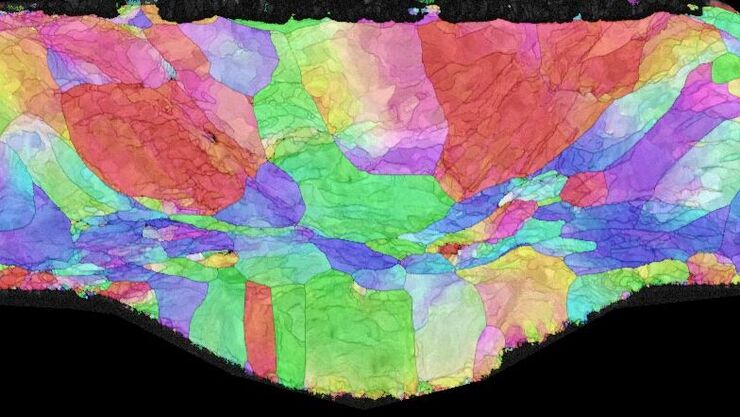
Introduction to Ion Beam Etching with the EM TIC 3X
In this article you can learn how to optimize the preparation quality of your samples by using the ion beam etching method with the EM TIC 3X ion beam milling machine. A short introduction of the…
Computational Clearing - Enhance 3D Specimen Imaging
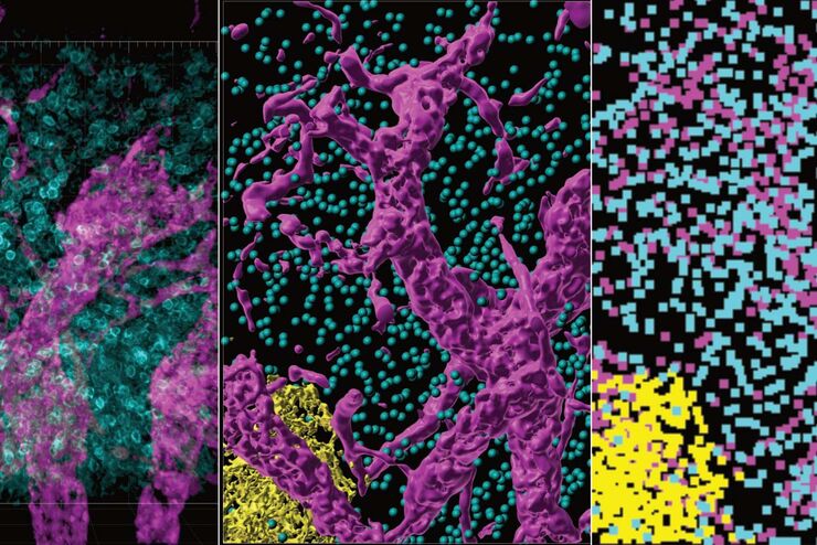
This webinar is designed to clarify crucial specifications that contribute to THUNDER Imagers' transformative visualization of 3D samples and improvements within a researcher's imaging-related…
STELLARIS White Light Lasers
When it comes to choosing fluorescent probes for your multi-color experiments, you shouldn’t have to compromise. Now you can advance beyond conventional excitation sources that limit your fluorophore…
Workflow Solutions for Sample Preparation Methods for Material Science
This brochure presents and explains appropriate workflow solutions for the most frequently required sample preparation methods for material science samples.
How to Drape an Overhead Surgical Microscope
The tutorial features the Leica ARveo digital Augmented Reality microscope for complex neurosurgery. The procedure also applies to the Leica M530 OHX, OH6, OH5 and OH4.
How to Drape a Surgical Microscope
Before performing surgical procedures, it is important to drape the surgical microscope to ensure sterile working conditions. At Leica, we are committed to helping you with your surgical practice. In…
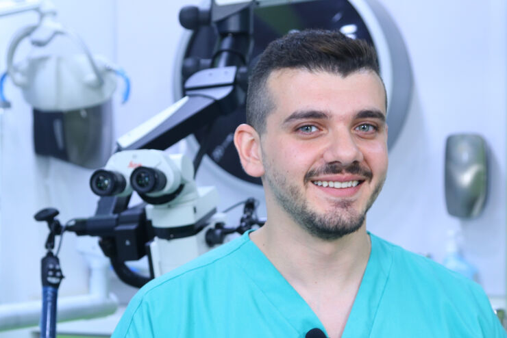
Overcoming Complexities in Microdentistry
Dr. Salam Abu Arqub, from the Smile Engineer Dental Center in Amman, Jordan, has been using Leica dental microscopes for three years for all procedures performed at the clinic. He shared his…
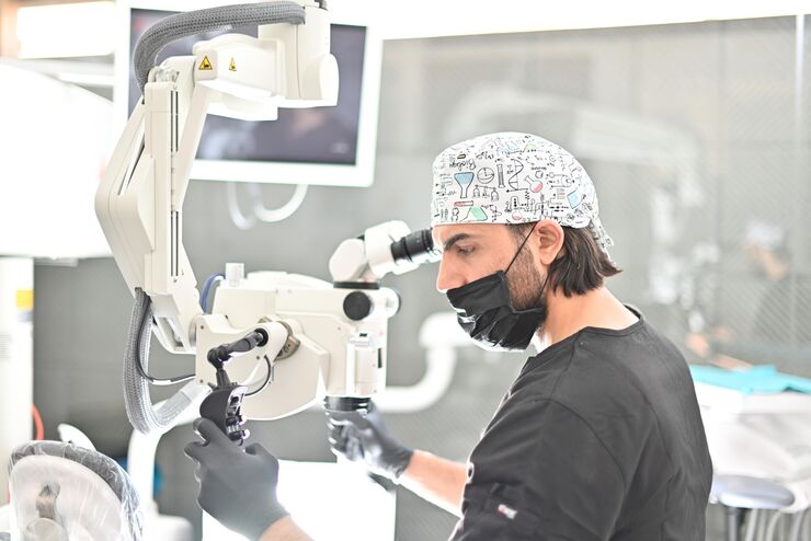
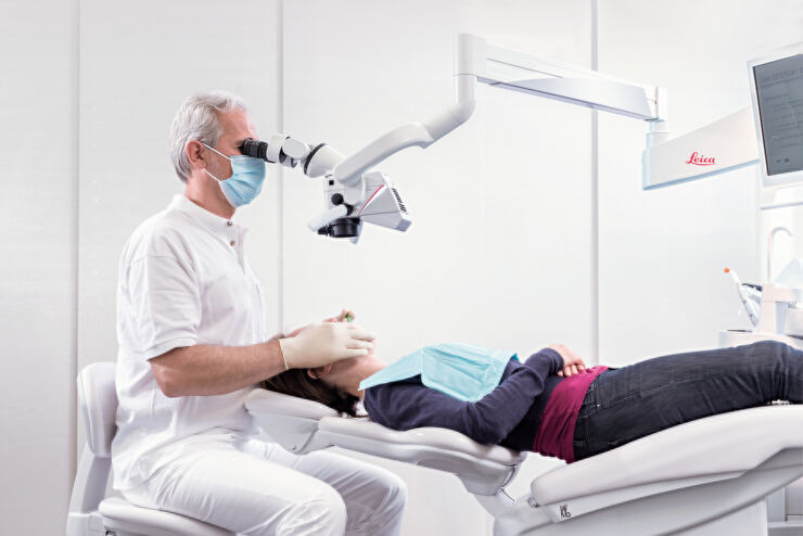
Minimally Invasive Dentistry: Visualization & Posture
Microscopes do not only provide better visualization during dental surgery. They also help ensure correct posture to avoid back pain and neck injuries. Dr. Iyad Ghoneim, from the Safad Dental Center…
THUNDER Imagers: High Performance, Versatility and Ease-of-Use for your Everyday Imaging Workflows
This webinar will showcase the versatility and performance of THUNDER Imagers in many different life science applications: from counting nuclei in retina sections and RNA molecules in cancer tissue…
Improve Cryo Electron Tomography Workflow
Leica Microsystems and Thermo Fisher Scientific have collaborated to create a fully integrated cryo-tomography workflow that responds to these research needs: Reveal cellular mechanisms at…
Improve 3D Cell Biology Workflow with Light Sheet Microscopy
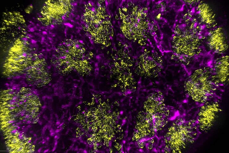
Understanding the sub-cellular mechanisms in carcinogenesis is of crucial importance for cancer treatment. Popular cellular models comprise cancer cells grown as monolayers. But this approach…
See More Than Just Your Image
Despite the emergence of new imaging methods in recent years, true 3D resolution is still achieved by Confocal Laser Scanning Microscopy (CLSM). Through a combination of novel, extremely fast scanning…
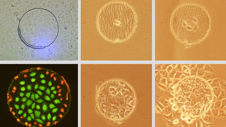
Live Cell Isolation by Laser Microdissection
Laser microdissection is a tool for the isolation of homogenous cell populations from their native niches in tissues to downstream molecular assays. Beside its routine use for fixed tissue sections,…
Successful Endodontic Treatment with Dental Operating Microscopes
In endodontics, accurate treatment is not only dependent on the technical skills and knowledge of the dentist, but also on clear, detailed visualization of the surgical field. As the outcome of an…
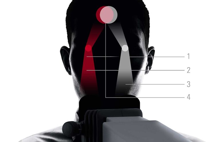
FusionOptics in Neurosurgery and Ophthalmology – for a Larger 3D Area in Focus
Neurosurgeons and ophthalmologists deal with delicate structures, deep or narow cavities and tiny structures with vitally important functions. A clear, three-dimensional view on the surgical field is…
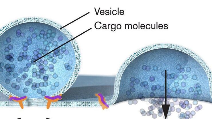
Nobel Prize 2013 in Physiology or Medicine for Discoveries of the Machinery Regulating Vesicle Traffic
On October 7th 2013, The Nobel Assembly at Karolinska Institutet has decided to award The Nobel Prize in Physiology or Medicine 2012 jointly to James E. Rothman, Randy W. Schekman and Thomas C. Südhof…

