Contact Us

James DeRose , Ph.D.

James DeRose

James DeRose is a Global Marketing Communication Manager at Leica Microsystems. His focus is on optimized content creation for applications concerning life science, material science, as well as industry and manufacturing. He is involved with application notes, case studies, technical reports, white papers, application pages, product pages, social media posts, emails, testimonials, and other materials. He has worked at Leica Microsystems since 2013. In the past, he worked on applications development projects in the fields of interfacial chemistry and physics, thermal and chemical engineering, corrosion and metallography, surface coatings, materials science, biotechnology, and cellular biology. He has expertise with various types of microscopy and analytical methods.
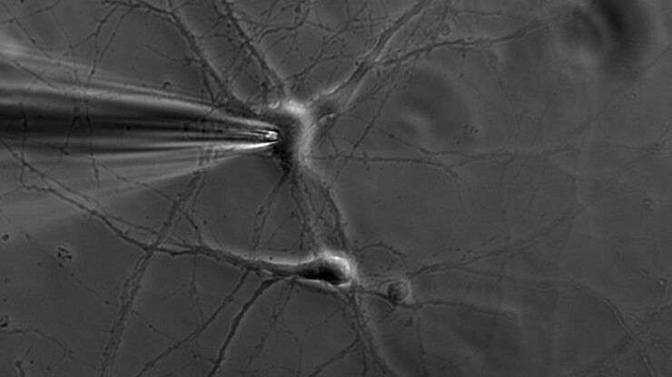
Patch pipette touching a murine hippocampal neuron. Image courtesy of A. Aguado, Ruhr University Bochum, Germany.

What is the Patch-Clamp Technique?

This article gives an introduction to the patch-clamp technique and how it is used to study the physiology of ion channels for neuroscience and other life-science fields.
Neurons imaged with DIC contrast.

Differential Interference Contrast (DIC) Microscopy

This article demonstrates how differential interference contrast (DIC) can be actually better than brightfield illumination when using microscopy to image unstained biological specimens.
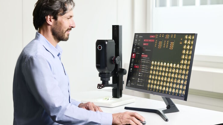
The Emspira 3 digital microscope offers what users need for comprehensive visual inspection, including comparison, measurement, and documentation sharing.

Digital Inspection Microscope for Industrial Applications

Factors users should consider before choosing a digital inspection microscope for industrial applications, including quality control (QC), failure analysis (FA), and R&D, are described in this…

Going Beyond Deconvolution

Widefield fluorescence microscopy is often used to visualize structures in life science specimens and obtain useful information. With the use of fluorescent proteins or dyes, discrete specimen…
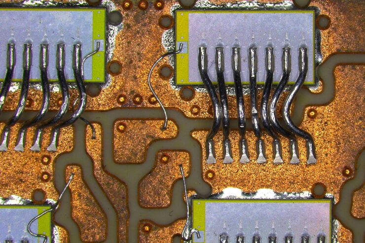
Inspection microscope image of a printed circuit board (PCB) taken with a ring light (RL) and near vertical illumination (NVI).

Microscope Illumination for Industrial Applications

Inspection microscope users can obtain information from this article which helps them choose the optimal microscope illumination or lighting system for inspection of parts or components.
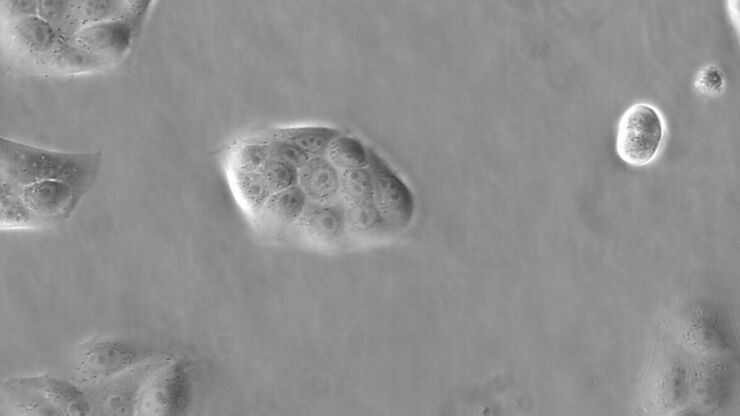
Image of MDCK (Madin-Darby canine kidney) cells taken with phase contrast.

Phase Contrast and Microscopy

This article explains phase contrast, an optical microscopy technique, which reveals fine details of unstained, transparent specimens that are difficult to see with common brightfield illumination.

Immersion Objectives

How an immersion objective, which has a liquid medium between it and the specimen being observed, helps increase the numerical aperture and microscope resolution is explained in this article.
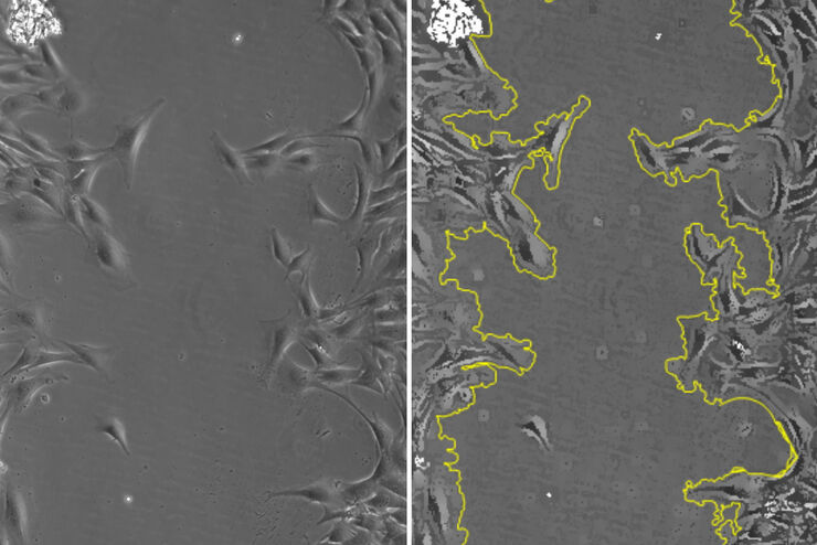
Phase-contrast image of a MDCK-cell culture and its respective confluency measured by the Mateo TL microscope.

How to Determine Cell Confluency with a Digital Microscope

This article shows how to measure cell confluency in an easy and consistent way with Mateo TL, increasing confidence in downstream experiments.
Intensity distribution (arbitrary color coding) of an image of two points where the distance between them corresponds to the Rayleigh criterion.

Microscope Resolution: Concepts, Factors and Calculation

This article explains in simple terms microscope resolution concepts, like the Airy disc, Abbe diffraction limit, Rayleigh criterion, and full width half max (FWHM). It also discusses the history.
Donor (D) and acceptor (A) molecule which participate in FRET (Förster resonance energy transfer).

What is FRET with FLIM (FLIM-FRET)?

This article explains the FLIM-FRET method which combines resonance energy transfer and fluorescence lifetime imaging to study protein-protein interactions.
Raw widefield and THUNDER image of transversal mouse adult fiber lens section. Courtesy N. Houssin, Plagemen lab, Ohio State University, Columbus, USA.

Studying Ocular Birth Defects

This article discusses how lens formation and ocular birth defects can be studied with sharp widefield microscopy images which are acquired rapidly. The mouse ocular lens is used as a model to study…
Automated Laser Microdissection for Proteome Analysis

Precise Spatial Proteomic Information in Tissues

Despite the availability of imaging-based and mass-spectrometry-based methods for spatial proteomics, a key challenge remains connecting images with single-cell-resolution protein abundance…
Image of murine-brain tissue showing a region removed with UV laser microdissection.

RNA Quality after Different Tissue Sample Preparation

The influence of sample preparation and ultraviolet (UV) laser microdissection (UV LMD) on the quality of RNA from murine-brain tissue cryo-sections is described in this article. To obtain good…
The various solutions from Leica Microsystems for cleanliness analysis.

Factors to Consider for a Cleanliness Analysis Solution

Choosing the right cleanliness analysis solution is important for optimal quality control. This article discusses the important factors that should be taken into account to find the solution that best…
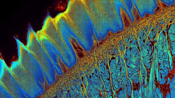
Transverse histological cut of a rabbit tongue. 50 Mpixels images (2326 µm x 1739 µm) in 14 x 18 tiles. Lifetime gives an additional contrast that allows to differentiate different structures in histological stainings.

A Guide to Fluorescence Lifetime Imaging Microscopy (FLIM)

The fluorescence lifetime is a measure of how long a fluorophore remains on average in its excited state before returning to the ground state by emitting a fluorescence photon.
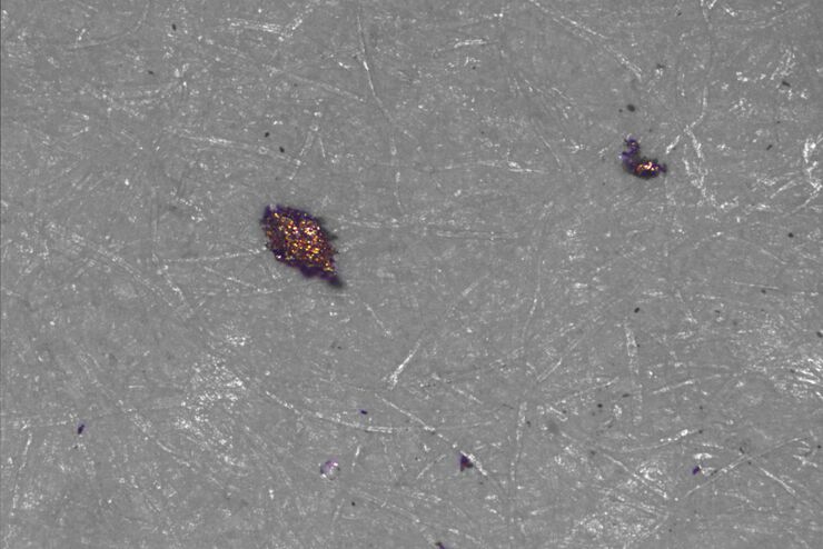
Type of contamination: spores

Cleanliness Analysis for Particulate Contamination

Devices, products, and their components fabricated in many industries can be quite sensitive to contamination and, as a result, have stringent requirements for technical cleanliness. Measurement…
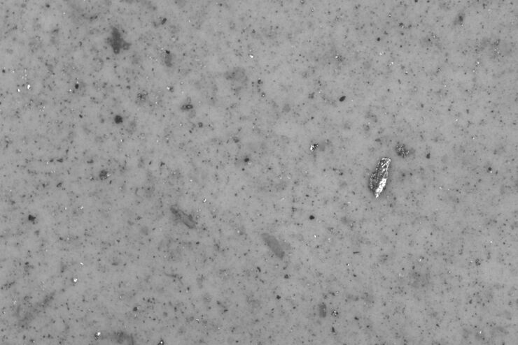
Particles and fibers on a filter which will be counted and analyzed for cleanliness

Efficient Particle Counting and Analysis

This report discusses particle counting and analysis using optical microscopy for cleanliness of parts and components. Particle counting and analysis is a critical part of quality assurance in the…
A stack of lithium-ion batteries

Quality Control Under the Microscope

Fast-rising demand for electric vehicles is one of the market’s main drivers, but there are other hotspots of growth, including the rise in renewable energy installations, such as photovoltaic panels,…
Scroll to top