Rolf T. Borlinghaus , Dr.

Rolf T. Borlinghaus

Rolf Borlinghaus was born 1956  in Grötzingen, Germany. After his diploma in Biology he worked on electrogenic steps of the Na/K-ATPase by laser-induced release of ATP from a caged compound at Peter Läuger’s Laboratory in the Biophysics Department, University Konstanz, Germany from where he was promoted to Dr.rer.nat. in 1988. He started working as a Product Manager for research Fluorescence and confocal Microscopes with Carl Zeiss, Oberkochen in 1990 and continued to tackle this challenge at Leica in 1997 (at that time Leica Lasertechnik, Heidelberg). For personal insights, in 2007, Rolf Borlinghaus dispensed his managerial responsibilities and is now supporting the confocal marketing group as senior scientist in a half-time position. The other half is dedicated to relations, food and botany.

Springer publications:

2017 Die Lichtblattmikroskopie (DE)

2017 The White Confocal (EN)

2016 Konfokale Mikroskopie in Weiß (DE)

2016 Unbegrenzte Lichtmikroskopie (DE)

Rolf Borlinghaus retired in December 2020.

After learning in May 2021 that Rolf has died, his colleagues would like to express their gratitude and appreciation for his numerous years of dedication to Leica Microsystems and its customers. He will be remembered for his many contributions to the field of microscopy which were published on Science Lab and in scientific journals and books.

Going Beyond Deconvolution

Widefield fluorescence microscopy is often used to visualize structures in life science specimens and obtain useful information. With the use of fluorescent proteins or dyes, discrete specimen…

Alzheimer Plaques: fast Visualization in Thick Sections

More than 60% of all diagnosed cases of dementia are attributed to Alzheimer’s disease. Typical of this disease are histological alterations in the brain tissue. So far, there is no cure for this…
Zebrafish Whole Brain imaging with Leica SP8 spectral confocal laser scanning microscope

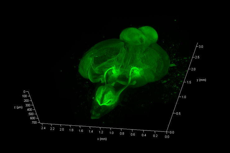
Zebrafish Brain - Whole Organ Imaging at High Resolution

Structural information is key when one seeks to understand complex biological systems, and one of the most complex biological structures is the vertebrate central nervous system. To image a complete…

What is a Resonant Scanner?

A resonant scanner is a type of galvanometric mirror scanner that allows fast image acquisition with single-point scanning microscopes (true confocal and multiphoton laser scanning). High acquisition…

What is a Spectral Detector (SP Detector)?

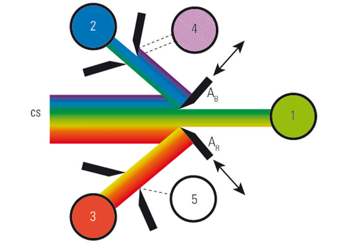
The SP detector from Leica Microsystems denotes a compound detection unit for point scanning microscopes, in particular confocal microscopes. The SP detector splits light into up to 5 spectral bands.…

Resolved Field Number (RFN)

The field number (FN) for optical microscopes indicates the field of view (FOV). It corresponds to the area in the intermediate image that is observable through the eyepieces. Although, we cannot…

What is a Field-of-View Scanner?

A field-of-view scanner is an assembly of galvanometric scanning mirrors used in single-point confocal microscopes that offer the correct optical recording of large field sizes. The field-of-view…

What is a Tandem Scanner?

A Tandem Scanner is an assembly of two different types of scanning together in one system for true confocal point scanning. The Tandem Scanner consists of a three-mirror scanning base with the…

Which Sensor is the Best for Confocal Imaging?

The Hybrid Photodetectors (HyD) are! Why that is the case is explained in this short Science Lab article.
Multiphoton microscopy of an unstained mouse skin section acquired using the 4Tune detector.

Mission Impossible Accomplished: Tunable Colors for Non-descanning Detection

Leica Microsystems’ 4Tune detector, the key component of the SP8 DIVE Deep In Vivo Explorer, provides spectrally tunable image recording with non-descanning detection. An innovative solution for…
„Confetti-Mouse“, taken with the 4Tune spectral detector of Leica’s Deep in-vivo Explorer SP8 DIVE.

Laser Beam Shaping for Multicolor Multiphoton Microscopy

Multiphoton Microscopy is one of the current hot topics in life science research. The new Leica TCS SP8 DIVE from Leica Microsystems presents a series of beneficial new innovations, including a freely…

Primary Beam Splitting Devices for Confocal Microscopes

Current fluorescence microscopy employs incident illumination which requires separation of illumination and emission light. The classical device performing this separation is a color-dependent beam…
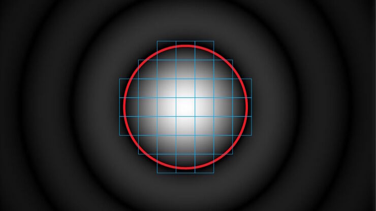
Pinhole diameter and diffraction pattern.

Pinhole Effect in Confocal Microscopes

When operating a confocal microscope, or when discussing features and parameters of such a device, we inescapably mention the pinhole and its diameter. This short introductory document is meant to…
© wowomnom – Fotolia.com

From Light to Mind: Sensors and Measuring Techniques in Confocal Microscopy

This article outlines the most important sensors used in confocal microscopy. By confocal microscopy, we mean "True Confocal Scanning", i.e. the technique that illuminates and measures one single…
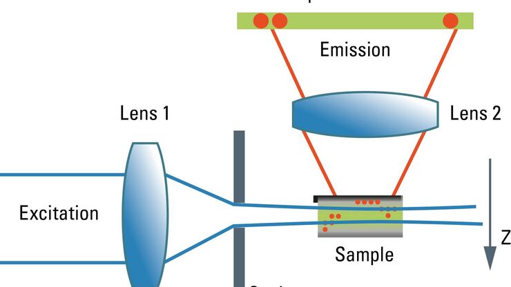
Schematic graph of the light path in a Spalt-Ultramikroskop.

Confocal and Light Sheet Imaging

Optical imaging instrumentation can magnify tiny objects, zoom in on distant stars and reveal details that are invisible to the naked eye. But it notoriously suffers from an annoying problem: the…
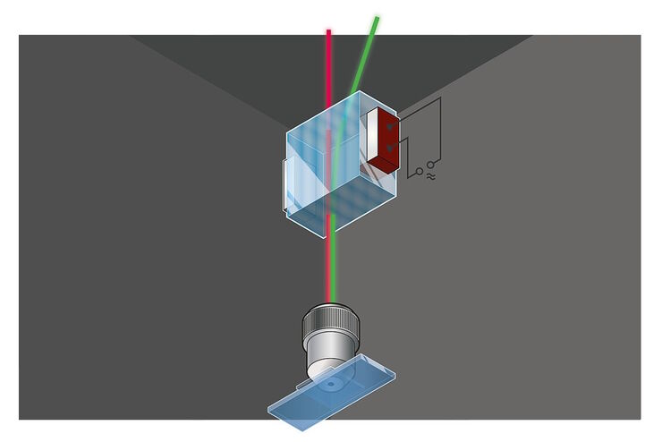
Acousto-optics, sketch

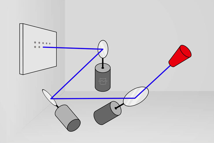
Acousto Optics in True Confocal Spectral Microscope Systems

Acousto-optical elements have successfully replaced planar filters in many positions. The white confocal, regarded as the fully spectrally tunable confocal microscope, was not possible without this…

Spectral Detection – How to Define the Spectral Bands that Collect Probe-specific Emission

To specifically collect emission from multiple probes, the light is first separated spatially and then passes through a device that defines a spectral band. Classically, this is a common glass-based…
Live cell imaging, 4 colors: Mitochondria (MitoView Green, yellow) and actin (mNeonGreen, cyan) microtubuli (SIR-tubulin, magenta), endosomes (NIR750, green). Processed with DSE and DSE powered by Aivia.

The Principles of White Light Laser Confocal Microscopy

The perfect light source for confocal microscopes in biomedical applications has sufficient intensity, tunable color and is pulsed for use in lifetime fluorescence. Furthermore, it should offer means…

Confocal Optical Section Thickness

Confocal microscopes are employed to optically slice comparably thick samples.
Scroll to top