
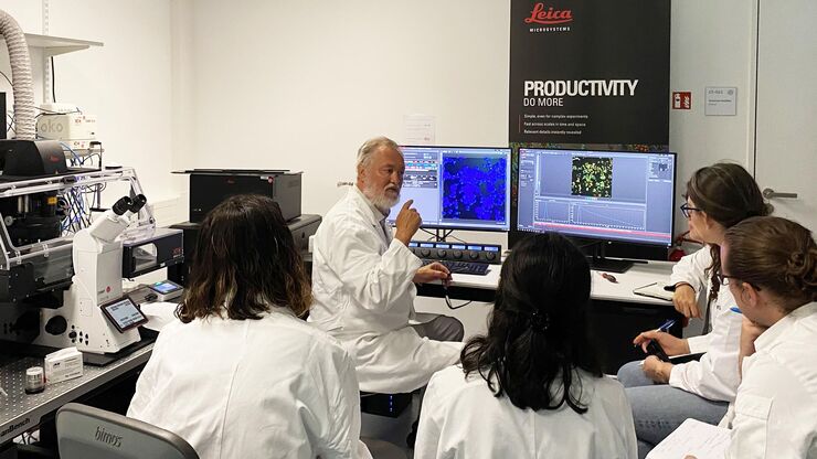
10-14 June 2023, Heidelberg, Germany – the course focusing on fluorescence lifetime-based readouts organized with the EMBL Imaging Centre gathered…
Leica and the EMBL Imaging Centre – Enabling Open Access
Throughout its history, Leica has enthusiastically developed relationships with academic and scientific research institutions to advance scientific understanding through microscopy. Now, thanks to our special partnership with the European Molecular Biology Laboratory (EMBL) in Heidelberg, researchers can gain access to cutting edge sample-prep and imaging technology.
By supporting EMBL’s drive to better understand the molecular basis of life through the provision of the latest technologies and expert support for a wide range of scientific and experimental services, Leica helps scientists push the boundaries of their research further and unlock greater insights.
Leica is among four industrial partners which helped to realize the vision of the EMBL Imaging Centre, which officially opened in 2022.
Find out more
Visit the EMBL IC website to see the instruments available to research applicants.
Latest Updates

The EMBO (European Molecular Biology Organization) practical course took place between the 12th and the 17th of February 2023. It covered all steps of…
 setting up the STELLARIS 8 STED for the virtual course")
Jointly organized by Leica Microsystems and EMBL, this virtual course held between the 8th and 15th of July 2022, provided attendees with deeper…
")
The EMBL Imaging Centre was inaugurated on the 30th of June 2022, welcoming representatives from politics, industry and research. The center was…

The EMBL in Heidelberg hosted its first Scientific Symposium at the EMBL Imaging Centre on the 30th of May 2022, featuring talks about leading edge…
Interviews
Interview with Yassin Harim, PhD Student at German Cancer Research Center, Heidelberg
Gain insights into 3D- whole mouse brain imaging using multicolor immunofluorescence! According to our guest user from the German Cancer Research Center, using THUNDER Imager Live cell at the EMBL IC was “…the perfect solution to get very high-quality images and also to spend little time on imaging because it's just so fast to acquire each individual slide”.
Interview with Virginia Pierini – Service Manager EMBL IC, Heidelberg. Virginia Pierini is supporting the operation of the EMBL Imaging Centre regarding all its services, with a special focus on users. She is the IC’s point of contact for all users, providing help around access procedures, the project execution as well as user training.
Interview with Virginia Pierini, Service Manager EMBL IC, Heidelberg
Virginia Pierini is supporting the operation of the EMBL Imaging Centre regarding all its services, with a special focus on users. She is the IC’s point of contact for all users, providing help around access procedures, the project execution as well as user training.
Interview with Giorgia Susin, PhD Student, University of Trento, Italy
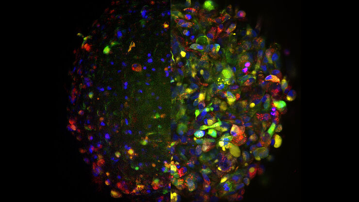
Giorgia Susin studies non-coding RNA transport in Xenopus laevis neurons using the STELLARIS microscope to unveil RNA-organelles interactions and enhance understanding of RNA transport. By performing live super-resolution imaging with TauSTED XTend for minimal light exposure, she preserved sample integrity while achieving high-resolution insights.
The EMBL Imaging Centre houses the latest state-of-the-art instrumentation from Leica and others, as well as those developed in EMBL research groups.
The EMBL IC offers researchers access to scientific experts from both academia as well as industry, providing its users with the opportunity to perform cutting-edge science with a suite of tools and support that are unavailable to most scientists.
Leica experts are on-site permanently at the EMBL IC to empower researchers to use the data from its advanced imaging systems in order to achieve groundbreaking insights.


Apply now
Bring your research project to EMBL and get support by the Leica Microsystems application specialists.
EMBL related articles

From Bench to Beam: A Complete Correlative Cryo Light Microscopy Workflow
In the webinar entitled "A Multimodal Vitreous Crusade, a Cryo Correlative Workflow from Bench to Beam" a team of experts discusses the exciting world of correlative workflows for structural biology…
Harnessing Microfluidics to Maintain Cell Health During Live-Cell Imaging
VIDEO ON DEMAND - In this webinar on-demand, we will use microfluidics to explore the effect of shear stress on cell morphology, examine the effect of nutrient replenishment on cellular growth during…
 and CellEvent™ (yellow).")
Following Multiple Events during Staurosporine Apoptosis
In this video on demand, we show how adding additional markers to an apoptosis kit can markedly increase the amount of information a researcher can obtain from the same experiment. The simultaneous…
, the cis-golgi matrix protein GM130 (AF488, green), and the trans-golgi network membrane protein TGN46 (AF647, red).")
Golgi Organizational Changes in Response to Cell Stress
In this video on demand, our special guest George Galea from EMBL Heidelberg will look at HeLa Kyoto cells treated with various chemotherapeutic agents to investigate their effect on the Golgi complex…

Imaging of Cardiac Tissue Regeneration in Zebrafish
Learn how to image cardiac tissue regeneration in zebrafish focusing on cell proliferation and response during recovery with Laura Peces-Barba Castaño from the Max Planck Institute.
How Does The Cytoskeleton Transport Molecules?
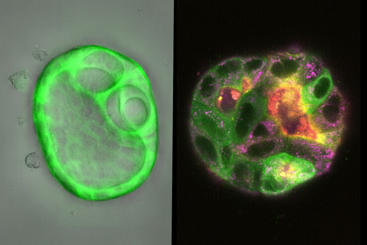
VIDEO ON DEMAND - See how 3D cysts derived from MDCK cells help scientists understand how proteins are transported and recycled in tissues and the role of the cytoskeleton in this transport.
 embryo, from sphere stage to somite stages.")
Studying Early Phase Development of Zebrafish Embryos
This video on demand focuses on combining widefield and confocal imaging to study the early-stage development of zebrafish embryos (Danio rerio), from oocyte to multicellular stage.

How To Get Multi Label Experiment Data With Full Spatiotemporal Correlation
This video on demand focuses on the special challenges of live cell experiments. Our hosts Lynne Turnbull and Oliver Schlicker use the example of studying the mitochondrial activity of live cells.…

How to Target Fluorescent Structures in 3D for Cryo-FIB Milling
This article describes the major steps of the cryo-electron tomography workflow including super-resolution cryo-confocal microscopy. We describe how subcellular structures can be precisely located in…

Benefits of TauContrast to Image Complex Samples
In this interview, Dr. Timo Zimmermann talks about his experience with the application of TauSense tools and their potential for the investigation of demanding samples such as thick samples or…
Meet the Leica Team at the EMBL IC

Robert Kirmse
Robert received his PhD from the DKFZ and University of Heidelberg. As post-doc, he worked on tumor cell invasion at BioQuant, Heidelberg and in cryo EM at the University of Colorado, Boulder. He joined Leica in 2019 as senior manager for sample preparation and site lead in Vienna. Since October 2022 he leads Leica’s EMBL IC team for Scientific Innovation.

Falco Krüger
Falco holds a PhD in Biology from the University of Heidelberg, Germany. Joining Leica in 2018 as an Advanced Workflow Specialist, he has since progressed to lead the team of Life Science Application Managers. Currently, Falco is also responsible for overseeing the OEM & Licensing Business Development at Leica Microsystems.

Andrea Mülter
Andrea joined Leica as product manager. After leading the global application team, she currently manages Leica’s knowledge program and strategic relations with leading scientists. She obtained her PhD at NIH with Jennifer Lippincott-Schwartz and then worked with Ursula Klingmüller at DKFZ in Heidelberg to study signal transduction by systems biology.
Andreia Pinto
Andreia worked as an electron microscopy specialist in Lisbon for 11 years. In 2019, she moved to London to finish her PhD and work in the fields of AI and Covid-19. Currently, she is an Advanced Workflow Specialist at Leica Microsystems and is based at the EMBL Imaging Centre in Heidelberg.
EMBL collaboration – a timeline

