How FusionOptics works
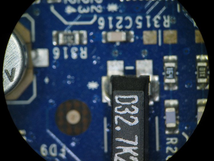
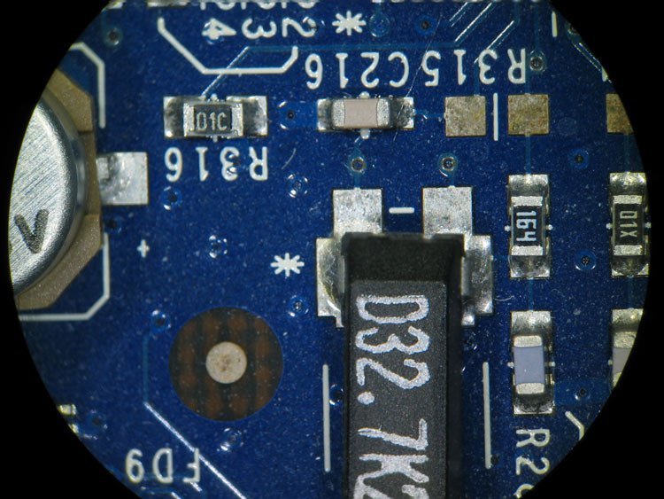
The FusionOptics principle makes use of the two separate beam paths of a stereo microscope, one beam for each eye. One beam path provides depth of field, the other provides high resolution. The human brain merges the two different images into a single, optimal 3D image.
Optimal 3D perception: The benefits of FusionOptics
Compared to conventional stereo microscopes, microscope users can perceive images with an up to three times larger depth of field as a result of FusionOptics. They need to refocus less, which saves them time, and they can stay focused on their observation task at hand – which is particularly helpful for detailed examination.
FusionOptics also holds immense benefits for inspection and rework in manufacturing, as users can see more with a greater depth of field and better estimate vertical distances for optimal results when they manipulate their samples with tools. The same advantages apply to microscopy work in the life sciences.
The image that FusionOptics produces also has a higher resolution than one from a conventional stereo microscope. Thus, users can see more fine details in the image – again a benefit for industrial, life-science, and medical applications alike.
The ingenious idea behind FusionOptics
The idea behind FusionOptics is based on scientific studies about visual perception and vision problems. These studies had shown that the brain can selectively process information from individual eyes and that it is very capable of compensating for differences in the visual acuity of the two eyes.
Breakthrough for stereo microscopes only
These studies uncovered an opportunity that can only be exploited with stereo microscopes, as they essentially work like an extension of our two eyes. They enable us to view microstructures in 3D with the help of two different beam paths which produce a separate image for each eye.
The catch when it comes to stereo microscopes is that ever since Horatio S. Greenough invented the first one, they work according to optical principles based primarily on Ernst Abbe’s work. After over a century, optics designers seemingly had pushed magnification, resolution and image quality to the limit permitted by optics. Once FusionOptics was invented, the situation changed.
Limits are made to be broken
These limits are determined by the correlation between resolution, convergence angle, and working distance. The higher the microscope resolution, the higher the convergence angle between the left and right beam paths and the lower the available working distance.
However, increasing the distance between the optical axes would cause the three-dimensional image seen by the observer to become distorted; a cube in the object would then appear as a tall tower. A greater zoom range alone is of little use, since with ever-increasing magnification, there would not always be an attendant increase in optical resolution. The result would be what is known as empty magnification [1].
The science behind FusionOptics
The above-mentioned studies gave the development engineers at Leica Microsystems the simple but ingenious idea: Why not take advantage of the brain’s ability and use each beam path of the microscope for different information? Thus, they found an innovative approach with FusionOptics by utilizing the human brain to do the work. The two advantages of increased resolution and higher depth of field can be achieved without increasing the convergence angle between the two beam paths.
This video explains FusionOptics in detail, showing the M205 FCA stereo microscope as an example.
Scientific study confirms FusionOptics
The feasibility of the FusionOptics design had to be reviewed in light of neurophysiology. A study tested whether the brain could process signals that differ in their local resolutions between the two eyes into correct three-dimensional images. Earlier studies were primarily concerned with two-dimensional images [2,3].
Leica Microsystems presented the idea to Dr. Daniel Kiper of the Institute of Neuroinformatics at the University of Zurich and Swiss Federal Institute of Technology [4], who specializes in research of signal processing in primate brains, and he agreed to carry out corresponding studies. Dr. Kiper, along with Graduate Assistant Cornelia Schulthess and Dr. Harald Schnitzler of Leica Microsystems, designed a study.
Testing the brain’s flexibility
A group of 36 subjects with normal visual acuity underwent psychophysical tests that investigated the binocular combination of visual signals. Of particular interest was whether an interocular signal suppression takes place when both eyes are exposed to different stimuli. The result of this test would be that the image of the suppressed eye would be perceived either only partially or not at all.
During the experiments, the test subjects observed patches arranged around a central fixation point. The fields either had patches with or without grids (Figure 1). To create differences in the perception of both eyes, binocular disparity is required – both eyes must be exposed to different stimuli. This was done using special stereo goggles with which separate test images could be projected to each eye.
In a series of trials, the test subjects saw changing arrangements of the grid patches in various planes at different depths compared to the plane of the central fixation point. After each image that was visible for 1,000 msec, the subjects reported where they saw the grid patches and whether they appeared in front of or behind the central fixation point.
Human brains select the best information from the eyes
The evaluation of the correct/incorrect answers for the position of the grid patches and the resolution in various planes showed no significant differences. No evidence of signal suppression was observed in any of the tests.
This result means that the human brain is capable of using the best information from both eyes in order to compose an optimal 3D image. This fact is true regardless of whether the same image is transmitted to the brain from both eyes or each eye provides entirely different information. The results prove once again how adaptable and powerful our brain is in processing visual impressions.
FusionOptics provides one-of-a-kind 3D images
On the theoretical basis provided by the study, Leica Microsystems was able to implement the FusionOptics concept in a stereo microscope with a large zoom range of 20.5:1 and a resolution of up to 1,050 line pairs (lp)/mm which corresponds to a visible structure width (a single line) of 476 nm. Up until some years ago, optical devices could only achieve a maximum zoom range of 16:1 or a magnification increase without an increase in resolution (empty magnification).
Breakthrough performance for several applications
The significant performance increase achieved by FusionOptics is valuable for everyday work at the microscope.
Whether for applications like inspection or quality control in the electronics, automotive, aerospace, or medical device industries, FusionOptics technology offers advantages that are not attainable with conventional stereo microscopy. Users doing applications relating to life sciences and medical research equally benefit from the performance, as Science Lab articles on developmental biology concerning zebrafish, medaka, and Xenopus illustrate [5,6]. And last, but not least, neurosurgeons and ophthalmologists, as well as their patients, benefit too, as fine details are resolved without the need to constantly refocus or change magnification [7].


Figure 3: Move the slider to see how FusionOptics combines larger depth of field with higher resolution to deliver an image of this PCB which is sharper and reveals more details than that of a conventional stereo microscope.


