
Industrial Microscopy
Industrial Microscopy
Dive deep into detailed articles and webinars focusing on efficient inspection, optimized workflows, and ergonomic comfort in industrial and pathological contexts. Topics covered include quality control, materials analysis, microscopy in pathology, among many others. This is the place where you get valuable insights into using cutting-edge technologies for improving precision and efficiency in manufacturing processes as well as accurate pathological diagnosis and research.
Filter articles
Tags
Products
Loading...

Brief Introduction to Coating Technology for Electron Microscopy
Coating of samples is required in the field of electron microscopy to enable or improve the imaging of samples. Creating a conductive layer of metal on the sample inhibits charging, reduces thermal…
Loading...
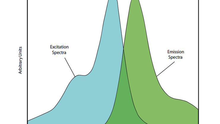
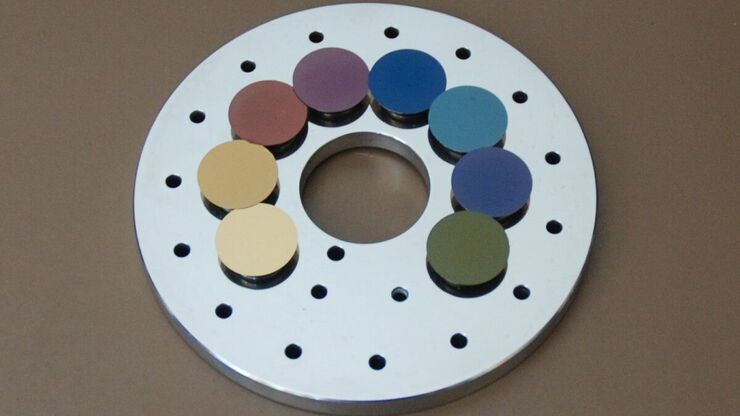
Handbook of Optical Filters for Fluorescence Microscopy
Fluorescence microscopy and other light-based applications require optical filters that have demanding spectral and physical characteristics. Often, these characteristics are application-specific and…
Loading...
Carbon Thickness Evaluation in Electron Microscopy
The coating layers applied and used for electron microscopy imaging are commonly controlled and measured by quartz crystals. These crystals oscillate with a certain frequency (around 6 megahertz when…
Loading...
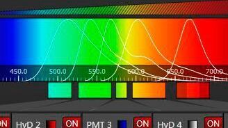
Spectral Detection – How to Define the Spectral Bands that Collect Probe-specific Emission
To specifically collect emission from multiple probes, the light is first separated spatially and then passes through a device that defines a spectral band. Classically, this is a common glass-based…
Loading...

Brief Introduction to High-Pressure Freezing
Water is the most abundant cellular constituent and therefore important for preserving cellular ultra-structure. Currently the only way to fix cellular constituents without introducing significant…
Loading...

Step by Step Guide for FRAP Experiments
Fluorescence Recovery After Photobleaching (FRAP) has been considered the most widely applied method for observing translational diffusion processes of macromolecules. The resulting information can be…
Loading...

Widefield Calcium Imaging with Calcium Indicator Fura2
In eukaryotic cells Ca2+ is one of the most widespread second messengers used in signal transduction pathways. Intracellular levels of Ca2+ are usually kept low, as Ca2+ often forms insoluble…
Loading...

Brief Introduction to Critical Point Drying
One of the uses of the Scanning Electron Microscope (SEM) is in the study of surface morphology in biological applications which requires the preservation of the surface details of a specimen. Samples…
Loading...

Nobel Prize 2012 in Physiology or Medicine for Stem Cell Research
The Nobel Prize recognizes two scientists who discovered that mature, specialised cells can be reprogrammed to become immature cells capable of developing into all tissues of the body. Their findings…
