
is mobile? false
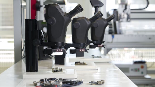
Industrial Microscopy
Industrial Microscopy
Dive deep into detailed articles and webinars focusing on efficient inspection, optimized workflows, and ergonomic comfort in industrial and pathological contexts. Topics covered include quality control, materials analysis, microscopy in pathology, among many others. This is the place where you get valuable insights into using cutting-edge technologies for improving precision and efficiency in manufacturing processes as well as accurate pathological diagnosis and research.
Popular Show subnavigation
Industrial Microscopy

Filter articles
Tags
Products
Loading...
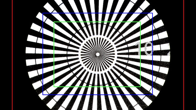
 which was imaged with extended depth of field (EDOF) using digital microscopy.")
Depth of Field in Microscope Images
For microscopy imaging, depth of field is an important parameter when needing sharp images of sample areas with structures having significant changes in depth. In practice, depth of field is…
Loading...

Deep Visual Proteomics Provides Precise Spatial Proteomic Information
Despite the availability of imaging methods and mass spectroscopy for spatial proteomics, a key challenge that remains is correlating images with single-cell resolution to protein-abundance…
Loading...

What is Empty Magnification and How can Users Avoid it
The phenomenon of “empty magnification”, which can occur while using an optical, light, or digital microscope, and how it can be avoided is explained in this article. The performance of an optical…
Loading...

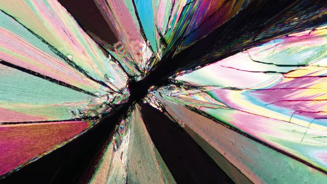
The Polarization Microscopy Principle
Polarization microscopy is routinely used in the material and earth sciences to identify materials and minerals on the basis of their characteristic refractive properties and colors. In biology,…
Loading...

Revealing Neuronal Migration’s Molecular Secrets
Different approaches can be used to investigate neuronal migration to their niche in the developing brain. In this webinar, experts from The University of Oxford present the microscopy tools and…
Loading...

Empowering Spatial Biology with Open Multiplexing and Cell DIVE
Spatial biology and multiplexed imaging workflows have become important in immuno-oncology research. Many researchers struggle with study efficiency, even with effective tools and protocols. Here, we…
Loading...

How did Laser Microdissection enable Pioneering Neuroscience Research?
Dr. Marta Paterlini, a Senior Scientist at the Karolinska Institute, shares her experience of using laser microdissection (LMD) in groundbreaking research into adult human neurogenesis and offers…
Loading...

How do Cells Talk to Each Other During Neurodevelopment?
Professor Silvia Capello presents her group’s research on cellular crosstalk in neurodevelopmental disorders, using models such as cerebral organoids and assembloids.
Loading...

Overcoming Observational Challenges in Organoid 3D Cell Culture
Learn how to overcome challenges in observing organoid growth. Read this article and discover new solutions for real-time monitoring which do not disturb the 3D structure of the organoids over time.
