
Industrial Microscopy
Industrial Microscopy
Dive deep into detailed articles and webinars focusing on efficient inspection, optimized workflows, and ergonomic comfort in industrial and pathological contexts. Topics covered include quality control, materials analysis, microscopy in pathology, among many others. This is the place where you get valuable insights into using cutting-edge technologies for improving precision and efficiency in manufacturing processes as well as accurate pathological diagnosis and research.
Filter articles
Tags
Story Type
Products
Loading...

Ratiometric Imaging
Many fundamental functions of a cell strongly depend on delicate, but nevertheless dynamic balances of ions (e.g. calcium, magnesium), voltage potentials and pH between the cell’s cytosol and the…
Loading...
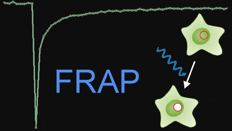
Fluorescence Recovery after Photobleaching (FRAP) and its Offspring
FRAP (Fluorescence recovery after photobleaching) can be used to study cellular protein dynamics: For visualization the protein of interest is fused to a fluorescent protein or a fluorescent dye. A…
Loading...

Förster Resonance Energy Transfer (FRET)
The Förster Resonance Energy Transfer (FRET) phenomenon offers techniques that allow studies of interactions in dimensions below the optical resolution limit. FRET describes the transfer of the energy…
Loading...
An Introduction to CARS Microscopy
CARS overcomes the drawbacks of conventional staining methods by the intrinsic characteristics of the method. CARS does not require labeling because it is highly specific to molecular compounds which…
Loading...
")
Mosaic Images
Confocal laser scanning microscopes are widely used to create highly resolved 3D images of cells, subcellular structures and even single molecules. Still, an increasing number of scientists are…
Loading...
Confocal Optical Section Thickness
Confocal microscopes are employed to optically slice comparably thick samples.
Loading...

Optical Contrast Methods
Optical contrast methods give the potential to easily examine living and colorless specimens. Different microscopic techniques aim to change phase shifts caused by the interaction of light with the…
Loading...

Integrated Modulation Contrast (IMC)
Hoffman modulation contrast has established itself as a standard for the observation of unstained, low-contrast biological specimens. The integration of the modulator in the beam path of themodern…
Loading...

Fluorescence in Microscopy
Fluorescence microscopy is a special form of light microscopy. It uses the ability of fluorochromes to emit light after being excited with light of a certain wavelength. Proteins of interest can be…
