Leica Microsystems

Leica Microsystems

Leica Microsystems is a world leader in microscopes and scientific instruments. Founded as a family business in the nineteenth century, the company’s history was marked by unparalleled innovation on its way to becoming a global enterprise.

Its historically close cooperation with the scientific community is the key to Leica Microsystems’ tradition of innovation, which draws on users’ ideas and creates solutions tailored to their requirements. At the global level, Leica Microsystems is organized in three divisions, all of which are among the leaders in their respective fields: Life Science, Industry and Medical.

The company is represented in over 100 countries with 6 manufacturing facilities in 5 countries, sales and service organizations in 20 countries, and an international network of dealers. The company is headquartered in Wetzlar, Germany.

http://www.leica-microsystems.com/

Fluorescence stereo microscope image of anesthetized Mediterranean fruit flies recorded with a M205 stereo microscope.

Investigating Fruit Flies (Drosophila melanogaster)

Learn how to image and investigate Drosophila fruit fly model organisms efficiently with a microscope for developmental biology applications from this article.
C. elegans

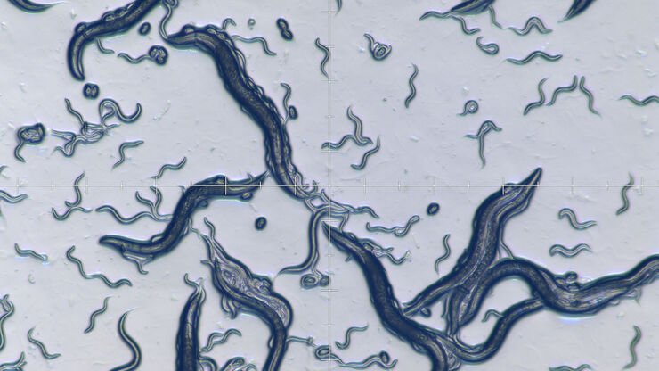
Studying Caenorhabditis elegans (C. elegans)

Find out how you can image and study C. elegans roundworm model organisms efficiently with a microscope for developmental biology applications from this article.

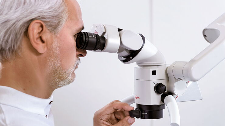
Six Features to Consider when Choosing a Dental Microscope

In dental medicine, the surgical microscope has become increasingly important for high-quality and successful surgeries, particularly in the field of endodontics. A microscope supports the dentist to…

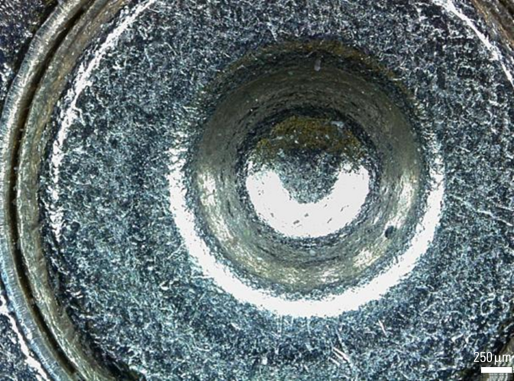
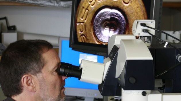
Digital Microscopy in Forensics

Forensic experts work with a broad range of microscopes to examine evidence from firearms and tool marks, documents, forensic or legal medicine, hair and fibers as well as glass and paint. Digital…
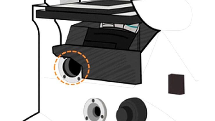
Infinity port

Infinity Optical Systems

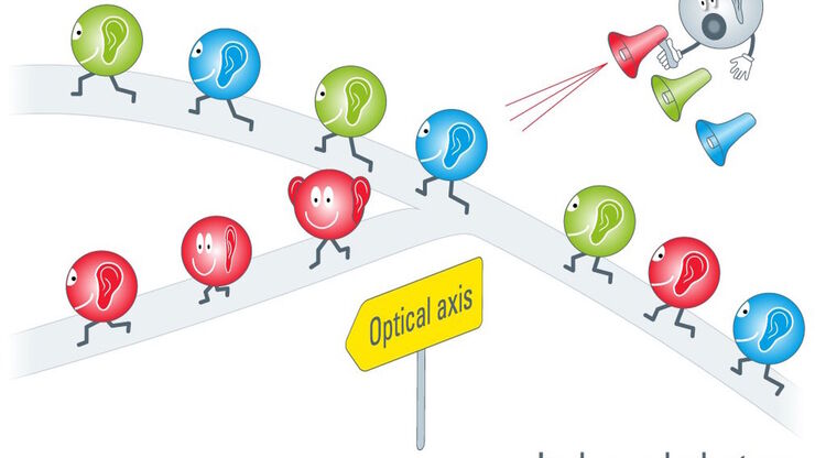
“Infinity Optics” refers to the concept of a beam path with parallel rays between the objective and the tube lens of a microscope. Flat optical components can be brought into this “Infinity Space”…

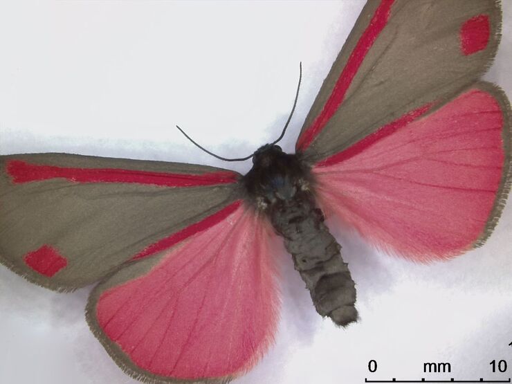
Life Science Imaging with DVM6 Digital Microscope

Digital microscopes can be a great help in life science applications such as the documentation in botany, entomology studies and crop science, or the digitization of museum collections. The image…
Proveo lightbeam

Cataract Surgery with CoAx4 Illumination

A stable red reflex is one of the most important features of an ophthalmic surgical microscope for cataract surgery. It’s the red reflex that makes the structure of the lens visible and thus makes for…

Workflows & Protocols: How to Isolate Individual Chromosomes with Laser Microdissection

During the first Leica Workshop in Brazil, at the Centro de Energia Nuclear na Agricultura/USP (CENA), the participants learned how to prepare samples for laser microdissection (LMD) using a cryotome.…
A 17th-century compound microscope (© Golub Collection – University of California, Berkeley/Steven Ruzin, Curator)

A Brief History of Light Microscopy

The history of microscopy begins in the Middle Ages. As far back as the 11th century, plano-convex lenses made of polished beryl were used in the Arab world as reading stones to magnify manuscripts.…
© wowomnom – Fotolia.com

From Light to Mind: Sensors and Measuring Techniques in Confocal Microscopy

This article outlines the most important sensors used in confocal microscopy. By confocal microscopy, we mean "True Confocal Scanning", i.e. the technique that illuminates and measures one single…

Workflows & Protocols: How to Use a Leica Laser Microdissection System and Qiagen Kits for Successful RNA Analysis

Laser Microdissection (LMD) allows isolating individual cells or chromosomes and is a well established technique for sample preparation prior downstream analysis of the nucleic acid content via PCR or…
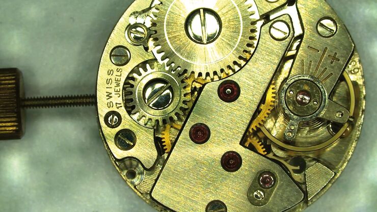
Watch imaged with DMS300.

What You Always Wanted to Know About Digital Microscopy, but Never Got Around to Asking

Digital microscopy is one of the buzz words in microscopy – and there are a couple of facts that are useful to know. Georg Schlaffer, Product Manager with Leica Microsystems, has often been asked…
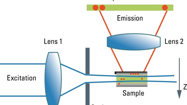
Schematic graph of the light path in a Spalt-Ultramikroskop.

Confocal and Light Sheet Imaging

Optical imaging instrumentation can magnify tiny objects, zoom in on distant stars and reveal details that are invisible to the naked eye. But it notoriously suffers from an annoying problem: the…

Universal PAINT – Dynamic Super-Resolution Microscopy

Super-resolution microscopy techniques have revolutionized biology for the last ten years. With their help cellular components can now be visualized at the size of a protein. Nevertheless, imaging…

Video Tutorials: Filling and Assembling of Different Carriers for High-Pressure Freezing

High pressure freezing (HPF) is a cryo-fixation method primarily for biological samples, but also for a variety of non-biological materials. It is a technique that yields optimal preservation in many…

FusionOptics in Neurosurgery and Ophthalmology – for a Larger 3D Area in Focus

Neurosurgeons and ophthalmologists deal with delicate structures, deep or narow cavities and tiny structures with vitally important functions. A clear, three-dimensional view on the surgical field is…

Brief Introduction to Freeze Fracture and Etching

Freeze fracture describes the technique of breaking a frozen specimen to reveal internal structures. Freeze etching is the sublimation of surface ice under vacuum to reveal details of the fractured…

Brief Introduction to Specimen Trimming

Before ultrathin sectioning a sample with an ultramicrotome it has to be pre-prepared. For this pre-preparation, special attention must be paid to the sample size (size of the section), location of…
Alexander von Inostranzeff (1843–1919)

125 Years of Comparison Microscopy

To be able to optically compare two objects with scientific accuracy, it must be possible to view them at the same time. This is particularly true for comparing small objects that can only be…

Sample Preparation for GSDIM Localization Microscopy – Protocols and Tips

The widefield super-resolution technique GSDIM (Ground State Depletion followed by individual molecule return) is a localization microscopy technique that is capable of resolving details as small as…
Acousto-optics, sketch

Acousto Optics in True Confocal Spectral Microscope Systems

Acousto-optical elements have successfully replaced planar filters in many positions. The white confocal, regarded as the fully spectrally tunable confocal microscope, was not possible without this…

Nobel Prize 2013 in Physiology or Medicine for Discoveries of the Machinery Regulating Vesicle Traffic

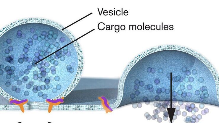
On October 7th 2013, The Nobel Assembly at Karolinska Institutet has decided to award The Nobel Prize in Physiology or Medicine 2012 jointly to James E. Rothman, Randy W. Schekman and Thomas C. Südhof…

Thermodynamic Considerations Regarding the LN2 in a High Pressure Freezer

Employing liquid nitrogen (LN2) as a coolant in the complex process of high pressure freezing raises certain considerations regarding phase transition not only of the liquid sample to be frozen but…
Lamellar structure

Brief Introduction to Contrasting for EM Sample Preparation

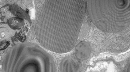
Since the contrast in the electron microscope depends primarily on the differences in the electron density of the organic molecules in the cell, the efficiency of a stain is determined by the atomic…

Brief Introduction to Glass Knifemaking for Electron and Light Microscope Applications

Glass knives are used in an ultramicrotome to cut ultrathin slices of samples for electron and light microscope applications. For resin and for cryo sections (Tokuyasu samples) the knife edge must be…

Brief Introduction to Coating Technology for Electron Microscopy

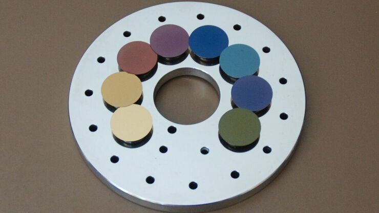
Coating of samples is required in the field of electron microscopy to enable or improve the imaging of samples. Creating a conductive layer of metal on the sample inhibits charging, reduces thermal…

Carbon Thickness Evaluation in Electron Microscopy

The coating layers applied and used for electron microscopy imaging are commonly controlled and measured by quartz crystals. These crystals oscillate with a certain frequency (around 6 megahertz when…

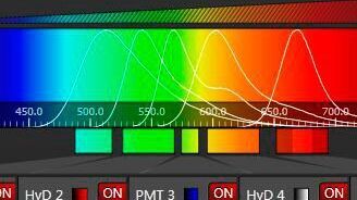
Spectral Detection – How to Define the Spectral Bands that Collect Probe-specific Emission

To specifically collect emission from multiple probes, the light is first separated spatially and then passes through a device that defines a spectral band. Classically, this is a common glass-based…
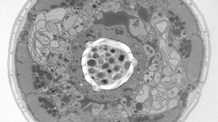
Structural details of the C. elegans, head in cross-section. Courtesy of Müller-Reichert T, MPI-CBG, Dresden, Germany, and McDonald K, University of California, Berkeley, USA.

Brief Introduction to High-Pressure Freezing

Water is the most abundant cellular constituent and therefore important for preserving cellular ultra-structure. Currently the only way to fix cellular constituents without introducing significant…

Step by Step Guide for FRAP Experiments

Fluorescence Recovery After Photobleaching (FRAP) has been considered the most widely applied method for observing translational diffusion processes of macromolecules. The resulting information can be…
Scroll to top