Contact Us

Leica Microsystems

Leica Microsystems

Leica Microsystems is a world leader in microscopes and scientific instruments. Founded as a family business in the nineteenth century, the company’s history was marked by unparalleled innovation on its way to becoming a global enterprise.

Its historically close cooperation with the scientific community is the key to Leica Microsystems’ tradition of innovation, which draws on users’ ideas and creates solutions tailored to their requirements. At the global level, Leica Microsystems is organized in three divisions, all of which are among the leaders in their respective fields: Life Science, Industry and Medical.

The company is represented in over 100 countries with 6 manufacturing facilities in 5 countries, sales and service organizations in 20 countries, and an international network of dealers. The company is headquartered in Wetzlar, Germany.
http://www.leica-microsystems.com/

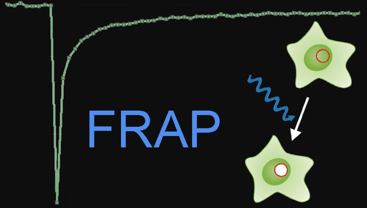
Step by Step Guide for FRAP Experiments

Fluorescence Recovery After Photobleaching (FRAP) has been considered the most widely applied method for observing translational diffusion processes of macromolecules. The resulting information can be…
Forensics microscopy

Every Clue Counts – Forensics Inconceivable Without Microscopy

There is no crime without clues. They may be obvious, like a cartridge case at the scene of the crime or clear signs of crowbar damage on a door. But sometimes, clues are microscopically small.…
Eukaryotic cells

Widefield Calcium Imaging with Calcium Indicator Fura2

In eukaryotic cells Ca2+ is one of the most widespread second messengers used in signal transduction pathways. Intracellular levels of Ca2+ are usually kept low, as Ca2+ often forms insoluble…
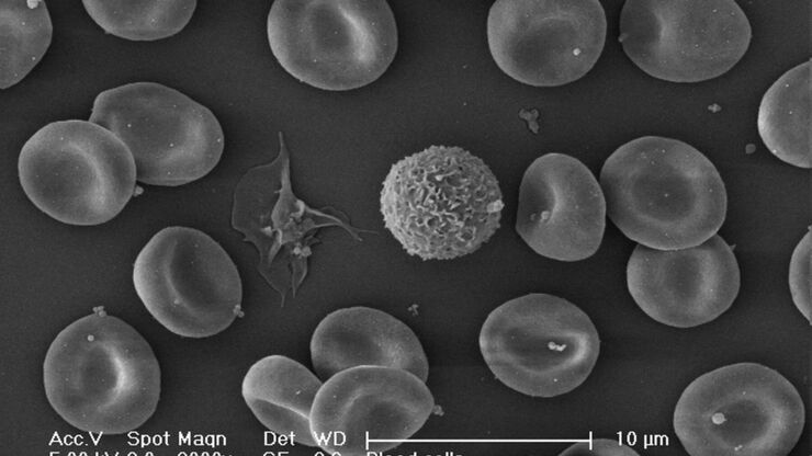
Blood cells. Critical Point Drying.

Brief Introduction to Critical Point Drying

One of the uses of the Scanning Electron Microscope (SEM) is in the study of surface morphology in biological applications which requires the preservation of the surface details of a specimen. Samples…
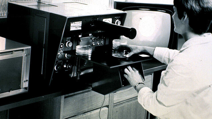
QTM B, 1963, the first commercial automated image analysis system for microscope images, based on a TV camera and developed by Metals Research in Cambridge, England.

50 Years of Image Analysis

Modern image analysis systems perform highly sophisticated image processing functions on images from an automated microscope and digital camera. 50 years ago, the first image analysis system was…
Center a fluorescence bulb.

Video Tutorial: How to Align the Bulb of a Fluorescence Lamp Housing

The traditional light source for fluorescence excitation is a fluorescence lamp housing with mercury burner. A prerequisite for achieving bright and homogeneous excitation is the correct centering and…
Exchange a fluorescence bulb.

Video Tutorial: How to Change the Bulb of a Fluorescence Lamp Housing

When applying fluorescence microscopy in biological applications, a lamp housing with mercury burner is the most common light source. This video tutorial shows how to change the bulb of a traditional…
Sub-Femtolitre volume_Fluorescence correlation spectroscopy (FCS)

Fluorescence Correlation Spectroscopy (FCS)

Fluorescence correlation spectroscopy ( FCS ) measures fluctuations of fluorescence intensity in a sub-femtolitre volume to detect such parameters as the diffusion time, number of molecules or dark…
White Light Laser: Lambda square scan

White Light Laser

The perfect light source for confocal microscopes in biomedical applications has sufficient intensity, tunable color and is pulsed for use in lifetime fluorescence. Furthermore, it should offer means…

Controlling the TIRF Penetration Depth is Mandatory for Reproducible Results

The main feature of total internal reflection fluorescence (TIRF) microscopy is the employment of an evanescent wave for the excitation of fluorophores instead of using direct light. A property of the…
Primary leaves of cowpea (Vigna unguiculata "California Blackeye") inoculated with cowpea mosaic virus (CPMV) containing the GFP-gene inserted between the movement protein (MP) and the capsid proteins (CPs) in the viral RNA 2

Introduction to Live-Cell Imaging

The understanding of complex and fast cellular dynamics is an important step to get insight into biological processes. Therefore, today’s life science research more and more demands studying…
TIRF Image of Tubulin, YFP, penetration depth: 120 mm

Applications of TIRF Microscopy in Life Science Research

The special feature of TIRF microscopy is the employment of an evanescent field for fluorophore excitation. Unlike standard widefield fluorescence illumination procedures with arc lamps, LEDs or…
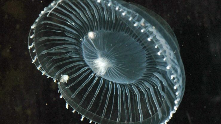
Jellyfish Aequorea Victoria

Fluorescent Proteins - From the Beginnings to the Nobel Prize

Fluorescent proteins are the fundament of recent fluorescence microscopy and its modern applications. Their discovery and consequent development was one of the most exciting innovations for life…
Snapshot from a time lapse of a calcium imaging experiment using the ratiometric calcium indicator Fura-2.

Ratiometric Imaging

Many fundamental functions of a cell strongly depend on delicate, but nevertheless dynamic balances of ions (e.g. calcium, magnesium), voltage potentials and pH between the cell’s cytosol and the…

Fluorescence Recovery after Photobleaching (FRAP) and its Offspring

FRAP (Fluorescence recovery after photobleaching) can be used to study cellular protein dynamics: For visualization the protein of interest is fused to a fluorescent protein or a fluorescent dye. A…

Förster Resonance Energy Transfer (FRET)

The Förster Resonance Energy Transfer (FRET) phenomenon offers techniques that allow studies of interactions in dimensions below the optical resolution limit. FRET describes the transfer of the energy…

An Introduction to CARS Microscopy

CARS overcomes the drawbacks of conventional staining methods by the intrinsic characteristics of the method. CARS does not require labeling because it is highly specific to molecular compounds which…
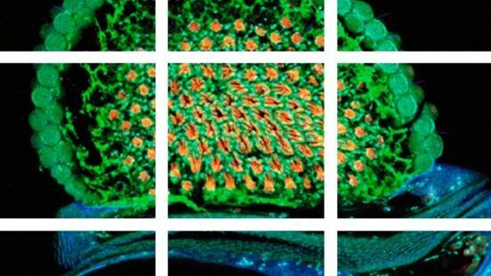
Scheme of a 2D mosaic scan. Drosophila melanogaster (eye section)

Mosaic Images

Confocal laser scanning microscopes are widely used to create highly resolved 3D images of cells, subcellular structures and even single molecules. Still, an increasing number of scientists are…
An ID card which has been tampered with by counterfeiters who inserted a hologram.

Is that Document Genuine or Fake? How do They Identify Fake Documents?

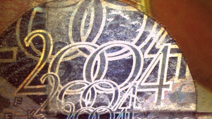
This article shows how forensic experts use microscopy for analysis to identify counterfeit, fake documents, such as ID cards, passports, visas, certificates, etc. Then they know if it is genuine or…

Confocal Optical Section Thickness

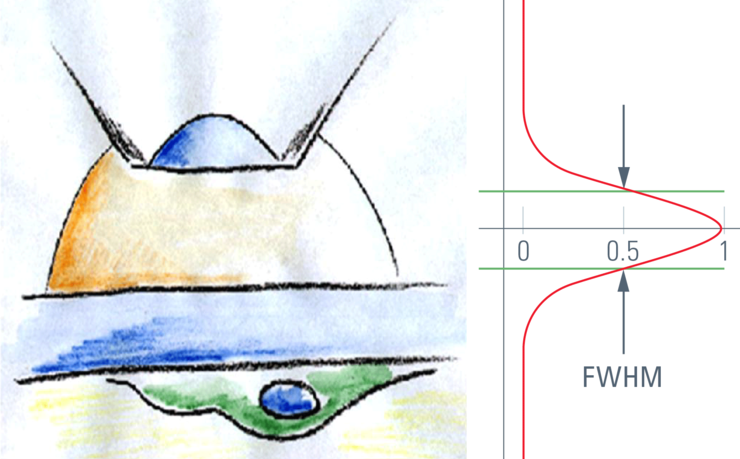
Confocal microscopes are employed to optically slice comparably thick samples.
Modulation contrast visualizes transparent, low-contrast specimens.

Integrated Modulation Contrast (IMC)

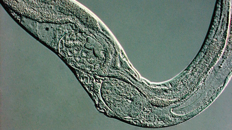
Hoffman modulation contrast has established itself as a standard for the observation of unstained, low-contrast biological specimens. The integration of the modulator in the beam path of themodern…
Object planes of the Greenough stereomicroscope with depth of field range.

Depth of Field in Microscopy

In microscopy, depth of field is often seen as an empirical parameter. In practice it is determined by the correlation between numerical aperture, resolution and magnification. For the best possible…
Cochlea implant. Illustration: © MED-EL.

Cochlea Implants for Deaf and Severely Hard of Hearing

The cochlea implant (CI) replaces the function of the outer ear, the middle ear and the ­cochlea of the inner ear. Basically, it consists of an external unit comprising a microphone, speech processor,…

New Standard in Electrophysiology and Deep Tissue Imaging

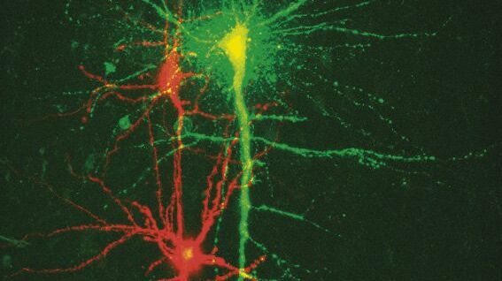
The function of nerve and muscle cells relies on ionic currents flowing through ion channels. These ion channels play a major role in cell physiology. One way to investigate ion channels is to use…
A portion of an early binocular microscope developed by John Leonhard Riddel in the early 1850s.

The History of Stereo Microscopy

This article gives an overview on the history of stereo microscopes. The development and evolution from handcrafted instruments (late 16th to mid-18th century) to mass produced ones the last 150…
Scroll to top