Contact Us

Leica Microsystems

Leica Microsystems

Leica Microsystems is a world leader in microscopes and scientific instruments. Founded as a family business in the nineteenth century, the company’s history was marked by unparalleled innovation on its way to becoming a global enterprise.

Its historically close cooperation with the scientific community is the key to Leica Microsystems’ tradition of innovation, which draws on users’ ideas and creates solutions tailored to their requirements. At the global level, Leica Microsystems is organized in three divisions, all of which are among the leaders in their respective fields: Life Science, Industry and Medical.

The company is represented in over 100 countries with 6 manufacturing facilities in 5 countries, sales and service organizations in 20 countries, and an international network of dealers. The company is headquartered in Wetzlar, Germany.

http://www.leica-microsystems.com/

See the Structure with Microscopy - Know the Composition with Laser Spectroscopy

The advantages of a 2-in-1 materials analysis solution combining optical microscopy and laser induced breakdown spectroscopy (LIBS) for simultaneous visual and chemical inspection are described in…

Visualization of DNA Molecules

Precise low angle rotary shadowing with heavy metals (like platinum) can be used in transmission electron microscopy (TEM) to observe molecular details of objects previously absorbed on a thin, low…

The Future of Fluorescence in Vascular Neurosurgery

In vascular neurosurgery, surgical microscopes are used to provide a magnified and illuminated view of the surgical field. Although surgeons benefit greatly from the superb image quality and optical…
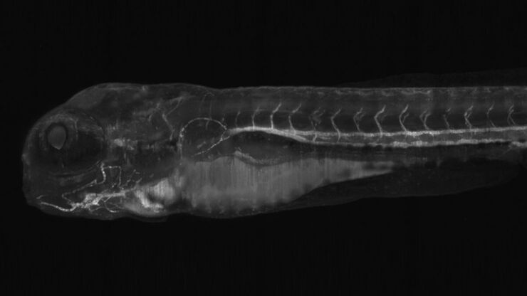
Light sheet microscopy tilescan of zebrafish.

Using U-Shaped Glass Capillaries for Sample Mounting

The DLS microscope system from Leica Microsystems is an innovative concept which integrates the Light Sheet Microscopy technology into the confocal platform. Due to its unique optical architecture,…

Practical Applications of Broad Ion Beam Milling

Mechanical polishing can be time consuming and frustrating. It can also introduce unwanted artifacts when preparing cross-sectioned samples for electron backscatter diffraction (EBSD) in the scanning…
Basalt imaged with crossed polarizers

Digital Microscopy in Earth Science

Classical polarized light (compound) microscopes can only be used for prepared samples, because the working distance they offer is insufficient for whole samples. This means that thicker and bigger…
Multiphoton microscopy of an unstained mouse skin section acquired using the 4Tune detector.

Mission Impossible Accomplished: Tunable Colors for Non-descanning Detection

Leica Microsystems’ 4Tune detector, the key component of the SP8 DIVE Deep In Vivo Explorer, provides spectrally tunable image recording with non-descanning detection. An innovative solution for…
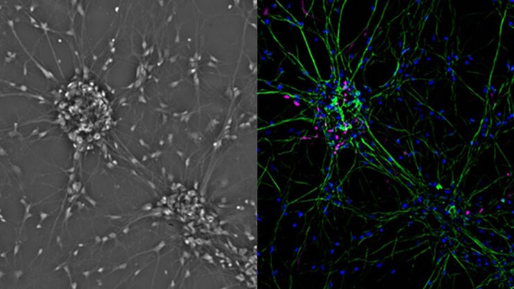
Mammalian cell culure. Phase contrast and fluorescence image.

Introduction to Mammalian Cell Culture

Mammalian cell culture is one of the basic pillars of life sciences. Without the ability to grow cells in the lab, the fast progress in disciplines like cell biology, immunology, or cancer research…
„Confetti-Mouse“, taken with the 4Tune spectral detector of Leica’s Deep in-vivo Explorer SP8 DIVE.

Laser Beam Shaping for Multicolor Multiphoton Microscopy

Multiphoton Microscopy is one of the current hot topics in life science research. The new Leica TCS SP8 DIVE from Leica Microsystems presents a series of beneficial new innovations, including a freely…
Convalaria

Introduction to Widefield Microscopy

This article gives an introduction to widefield microscopy, one of the most basic and commonly used microscopy techniques. It also shows the basic differences between widefield and confocal…

Optimization of the Interplay of Optical Components for Aberration Free Microscopy

Optical microscopes are used to magnify objects which are otherwise invisible for the human eye. For this purpose high quality optics is necessary to achieve appropriate resolution. However, besides…

Primary Beam Splitting Devices for Confocal Microscopes

Current fluorescence microscopy employs incident illumination which requires separation of illumination and emission light. The classical device performing this separation is a color-dependent beam…
Pinhole diameter and diffraction pattern.

Pinhole Effect in Confocal Microscopes

When operating a confocal microscope, or when discussing features and parameters of such a device, we inescapably mention the pinhole and its diameter. This short introductory document is meant to…
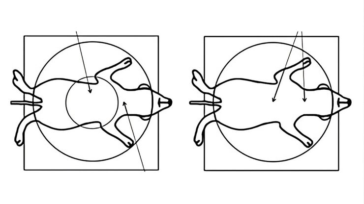
Comparison when observing a rodent model organism with a Greenough versus CMO (common main objective) stereo microscope for a task like surgery.

Rodent and Small-Animal Surgery

Learn how you can perform rodent (mouse, rat, hamster) and small-animal surgery efficiently with a microscope for developmental biology and medical research applications by reading this article.
Image Guided Surgery

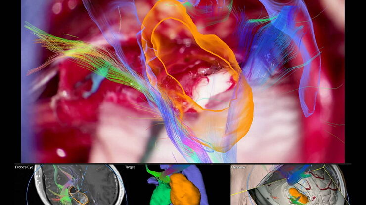
Navigating Through the Brain

One of the challenges of neurosurgery is orientation at the surgical site. When resecting tumors, removing arteriovenous malformations, or clipping aneurysms, surgeons often have to work near healthy…
Left: Tissue cells marked with an immunolabel (FITC) illuminated with wide-band UV excitation. Note the tissue structure with blue autofluorescence. Right: Same tissue and same immunostaining with FITC label illuminated with epi-illumination using narrow-band blue (490 nm) light. Note the increased image contrast (Ploem, 1967)

Milestones in Incident Light Fluorescence Microscopy

Since the middle of the last century, fluorescence microscopy developed into a bio scientific tool with one of the biggest impacts on our understanding of life. Watching cells and proteins with the…

Porous Ceramics - Sample Preparation for SEM

Application Note for Leica EM RES102 - Ceramic membrane filters with pore sizes down to a few nanometres must be investigated in cross-section with regard to the structure of the pores. The smallest…
Coaxial light with only polarizer open.

Digital Microscopy with Versatile Illumination and Various Contrast Methods for More Efficient Inspection and Quality Control

State-of-the-art digital microscopes utilizing a versatile illumination system capable of achieving multiple contrast methods, such as the Leica DVM6, are very useful for inspection, quality control,…
Scroll to top