Leica Microsystems

Leica Microsystems is a world leader in microscopes and scientific instruments. Founded as a family business in the nineteenth century, the company’s history was marked by unparalleled innovation on its way to becoming a global enterprise.
Its historically close cooperation with the scientific community is the key to Leica Microsystems’ tradition of innovation, which draws on users’ ideas and creates solutions tailored to their requirements. At the global level, Leica Microsystems is organized in three divisions, all of which are among the leaders in their respective fields: Life Science, Industry and Medical.
The company is represented in over 100 countries with 6 manufacturing facilities in 5 countries, sales and service organizations in 20 countries, and an international network of dealers. The company is headquartered in Wetzlar, Germany.


 taken with a ring light (RL) and near vertical illumination (NVI).")
 cells taken with phase contrast.")


 of an image of two points where the distance between them corresponds to the Rayleigh criterion.")


 and acceptor (A) molecule which participate in FRET (Förster resonance energy transfer).")
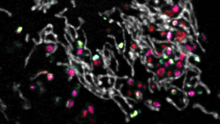
, SPY-Actin (cyan), and SiR-Tubulin (magenta). Instant Computational Clearing (ICC) was applied.")


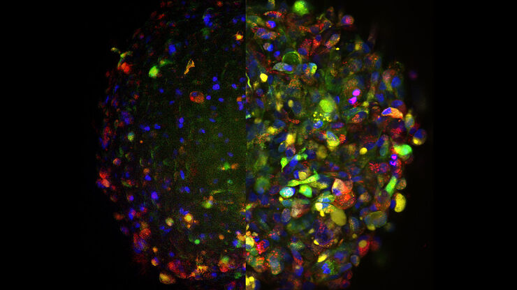
 and CellEvent™ (yellow).")


