Contact Us

Leica Microsystems

Leica Microsystems

Leica Microsystems is a world leader in microscopes and scientific instruments. Founded as a family business in the nineteenth century, the company’s history was marked by unparalleled innovation on its way to becoming a global enterprise.

Its historically close cooperation with the scientific community is the key to Leica Microsystems’ tradition of innovation, which draws on users’ ideas and creates solutions tailored to their requirements. At the global level, Leica Microsystems is organized in three divisions, all of which are among the leaders in their respective fields: Life Science, Industry and Medical.

The company is represented in over 100 countries with 6 manufacturing facilities in 5 countries, sales and service organizations in 20 countries, and an international network of dealers. The company is headquartered in Wetzlar, Germany.

http://www.leica-microsystems.com/

How to Select a Microscope for Cataract Surgery

What to consider in the selection of an ophthalmic microscope for cataract procedures. Bearing these aspects in mind will equip surgeons well for talks with manufacturer representatives. Many…
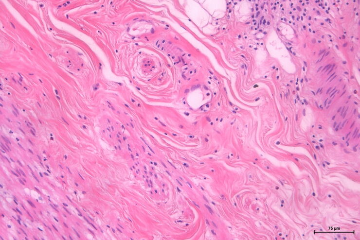
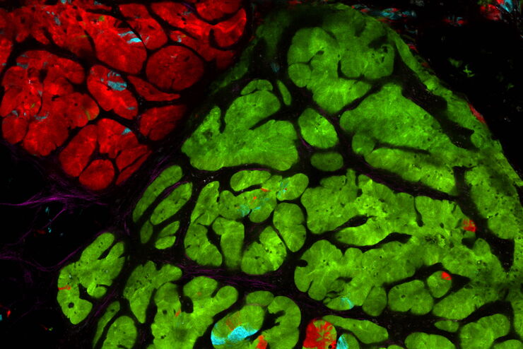
Spontaneous colon adenoma

Intravital Microscopy of Cancer

Join our guest speaker Prof Dr Jacco van Rheenen, as he presents his work on the identity, behavior and fate of cells that drive the initiation and progression of cancer.
Aivia_Neuroscience-VBE comparison mouse-1_traced_ROI

Accelerating Neuron Image Analysis with Automation

The ability to examine complex neural processes relies on the accurate reconstruction of neuronal networks at scale. Most data extraction methods in neuroscience research are time-consuming and…

Save Time and Effort with AI-assisted Fluorescence Image Analysis

The powerful synergy of THUNDER and Aivia analyze fluorescence images with greater accuracy, even when using low light excitation.
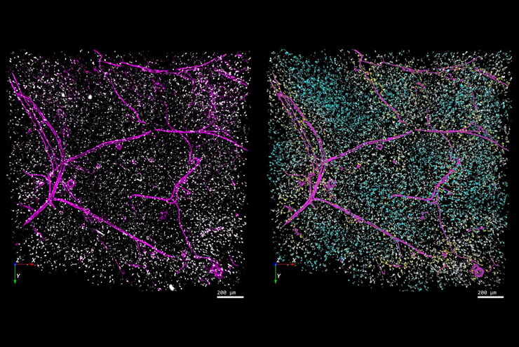
Separation of cells based on their tracking status: A colourised binary mask of a time-lapse microscopy field of view of medium confluency with individual cells highlighted as survivors if they can be tracked since the initial movie frame (cyan), incomers if they migrated into the field of view throughout the movie (yellow) or mistracks if an error occurred in the automated trajectory reconstruction (red).

Tracking Single Cells Using Deep Learning

AI-based solutions continue to gain ground in the field of microscopy. From automated object classification to virtual staining, machine and deep learning technologies are powering scientific…
Analysis of anatomy and axon orientation of an adult mouse brain tissue with QLIPP.

Learning the Cellular Architecture from its Optical Properties

In the last 3 years, microscopists have started to use "AI based" solutions for a wide range of applications, including image acquisition optimization (smart microscopy), object classification, image…
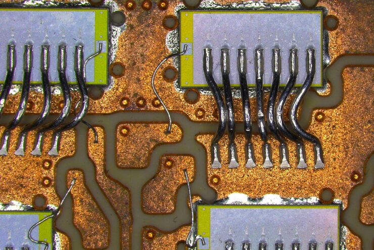
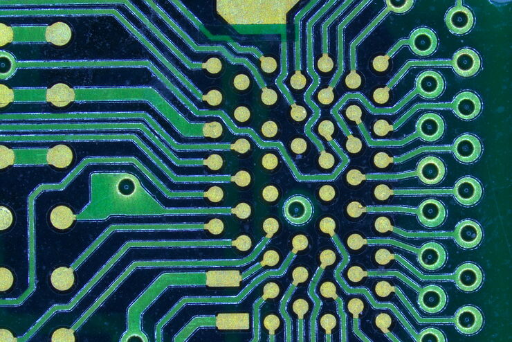
Visual inspection of a PCBA with the Ivesta 3 Greenough stereo microscope.

How to Select the Right Solution for Visual Inspection

This article helps users with the decision-making process when selecting a microscope as a solution for routine visual inspection. Important factors that should be considered are described.

How to Use a Digital Microscope to Streamline Inspection Processes

Watch this webinar for inspiration and expert advice on how to make quality control simpler, quicker, and easier. Learn how to perform comprehensive visual inspection, including comparison,…
Dynamic Signal Enhancement powered by Aivia:  Truly simultaneous multicolor imaging of live cells (U2OS) in 3D

Artificial Intelligence and Confocal Microscopy – What You Need to Know

This list of frequently asked questions provides “hands-on” answers and is a supplement to the introductory article about Dynamic Signal Enhancement powered by Aivia "How Artificial Intelligence…
Dynamic Signal Enhancement powered by Aivia: Truly simultaneous multicolor imaging of live cells (U2OS) in 3D

How Artificial Intelligence Enhances Confocal Imaging

In this article, we show how artificial intelligence (AI) can enhance your imaging experiments. Namely, how Dynamic Signal Enhancement powered by Aivia improves image quality while capturing the…

Designing your Research Study with Multiplexed IF Imaging

Multiplexed tissue analysis is a powerful technique that allows comparisons of cell-type locations and cell-type interactions within a single fixed tissue sample. It is common for researchers to ask…
SEM image of the full Li-NMC electrode sample, showing the two porous layers and the metal film at the center of the structure.

Cross Section Ion Beam Milling of Battery Components

Sample Preparation of Lithium battery systems requires high quality surface preparation to evaluate their internal structure and morphology. Due to the brittle materials involved, preparing pristine…

How to Successfully Perform Live-cell CLEM

The Leica Nano workflow provides a streamlined live-cell CLEM solution for getting insight bout structural changes of cellular components over time. Besides the technical handling described in the…

How to Successfully Implement Coral Life

The live-cell  CLEM workflow allows you to capture dynamic information related to a relevant biological process as it happens and put these observations into their ultrastructural context. The Leica…

Advancing Cellular Ultrastructure Research

Freeze-fracture and freeze-etching are useful tools for studying flexible membrane-associated structures such as tight junctions or the enteric glycocalyx. Freeze-fracture and etching are two…

Be Confident in your Results with Cell DIVE Validated Antibodies

The Cell DIVE System includes a carefully curated list of hundreds of commercially available antibodies validated to offer optimal specificity and sensitivity in multiplexed imaging. That validation…

Optimal Visualization in Brain Surgery

This case study “Treatment of the Galassi type III arachnoid cyst with the M530 OHX surgical microscope from Leica Microsystems” documents the procedure step by step and shows the visualization…

Benefits of Combining STED and Lifetime

In this interview, Professor Alberto Diaspro talks about the advantages of the White Light Laser and the TauSTED capabilities of STELLARIS 8 STED. He speaks about his experience with the confocal…

Spectroscopic Evaluation of Red Blood Cells

Hemoglobinopathies are a major healthcare problem. This study presents a possible diagnostic tool for thalassemia which is based on confocal spectroscopy. This approach exploits spectral detection and…

How to use a Surgical Microscope as an Operating Room Nurse

Surgical microscopes play an essential role in the modern microsurgery procedures. It provides the surgeon, assistant and operating room staff with a magnified and illuminated high-quality image of…
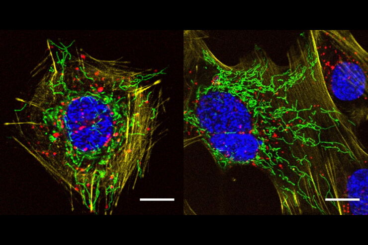
HeLa Kyoto cells (HKF1, H2B-mCherry, alpha Tubulin, mEGFP). Left image: Maximum projection of a z-stack prior to ICC and LVCC. Right image: Maximum projection of a mosaic z-stack after ICC and LVCC.

How to Improve Live Cell Imaging with Coral Life

For live-cell CLEM applications, light microscopy imaging is a critical step for identifying the right cell in the right state at the right time. In this article, Leica experts share their insights on…

The Cryo-CLEM Journey

This article describes the Cryo-CLEM technology and the benefits it can provide for scientists. Additionally, some scientific publications are highlighted. Recent developments in cryo electron…

Clinical Symposium on OCT-guided Cornea Surgery

In this recording Prof. Mehta from Singapore National Eye Centre and Prof. Fontana from Santa Maria Nuova Hospital in Regio Emilia, Italy, share their expertise on corneal surgery. They present PK,…
The EM ICE Nano loading area

How to Keep Your Samples Under Physiological Conditions

The Coral Life workflow combines dynamic data with the best possible sample fixation by high pressure freezing. However, good sample preservation won’t help if your cells are stressed by temperature…

Fluorescence Lifetime-based Imaging Gallery

Confocal microscopy relies on the effective excitation of fluorescence probes and the efficient collection of photons emitted from the fluorescence process. One aspect of fluorescence is the emission…

Optimizing THUNDER Platform for High-Content Slide Scanning

With rising demand for full-tissue imaging and the need for FL signal quantitation in diverse biological specimens, the limits on HC imaging technology are tested, while user trainability and…

Visualizing Protein Degradation and Aggregation in the Living Cell

Our guest speaker, Prof Dr Eric Reits, presents his work on neurodegenerative disorders. Reits’ group are experts on the subject of Huntington’s disease and work towards identifying leads for…

Towards Advanced Use of Intraoperative OCT in Cataract Surgery

In this White Paper, Dr. Rachid Tahiri shares his personal experience with the Leica EnFocus intraoperative OCT, the valuable features supporting smooth surgery and how it allows him to minimize…

How Clinical and Surgical Microscopes Support Otolaryngology

The number of ENT surgery procedures is increasing year after year. It is estimated that there will be over 21 million ENT procedures performed annually worldwide by 2022. Dr. Duane Mol is the…

Capture life as it happens

With the Leica Nano Workflow, searching for the needle in the haystack is a thing of the past. Take advantage of correlative light and electron microscopy to identify directly the right cell at the…
Scroll to top