Leica Microsystems

Leica Microsystems

Leica Microsystems is a world leader in microscopes and scientific instruments. Founded as a family business in the nineteenth century, the company’s history was marked by unparalleled innovation on its way to becoming a global enterprise.

Its historically close cooperation with the scientific community is the key to Leica Microsystems’ tradition of innovation, which draws on users’ ideas and creates solutions tailored to their requirements. At the global level, Leica Microsystems is organized in three divisions, all of which are among the leaders in their respective fields: Life Science, Industry and Medical.

The company is represented in over 100 countries with 6 manufacturing facilities in 5 countries, sales and service organizations in 20 countries, and an international network of dealers. The company is headquartered in Wetzlar, Germany.

http://www.leica-microsystems.com/

Visualizing Protein Degradation and Aggregation in the Living Cell

Our guest speaker, Prof Dr Eric Reits, presents his work on neurodegenerative disorders. Reits’ group are experts on the subject of Huntington’s disease and work towards identifying leads for…

Towards Advanced Use of Intraoperative OCT in Cataract Surgery

In this White Paper, Dr. Rachid Tahiri shares his personal experience with the Leica EnFocus intraoperative OCT, the valuable features supporting smooth surgery and how it allows him to minimize…

How Clinical and Surgical Microscopes Support Otolaryngology

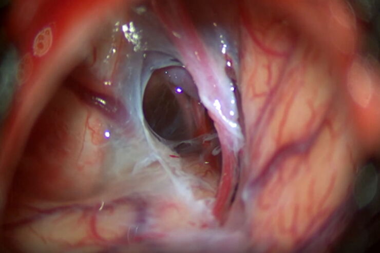
The number of ENT surgery procedures is increasing year after year. It is estimated that there will be over 21 million ENT procedures performed annually worldwide by 2022. Dr. Duane Mol is the…

Capture life as it happens

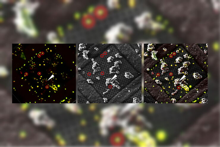
With the Leica Nano Workflow, searching for the needle in the haystack is a thing of the past. Take advantage of correlative light and electron microscopy to identify directly the right cell at the…

Microstructural Characterization including Compositional Analysis

Leica Microsystems' versatile upright compound microscope, DM6 M, fitted with Laser-Induced Breakdown Spectroscopy module will let you not only analyze metallographically polished samples and conduct…

Life Beyond the Pixels: Deep Learning Methods for Single Cell Analysis

Our guest speaker Prof Dr Peter Horvath presents his work on single cell-based large-scale microscopy experiments. This novel targeting approach includes the use of machine learning models and…

Introduction to 21 CFR Part 11 and Related Regulations

This article provides an overview of regulations and guidelines for electronic records (data entry, storage, signatures, and approvals) used in the USA (21 CFR Part 11), EU (GMP Annex 11), and China…
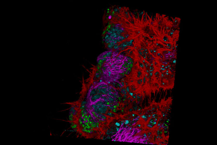
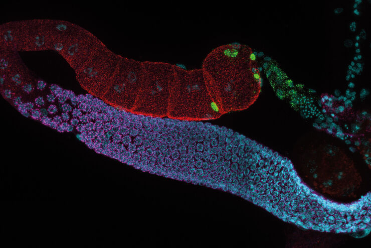
C. elegans Gonades - THUNDER Imager  Adult hermaphrodit, Staining: blue - DAPI (Nucleus), green - SP56 (sperms), red - RME-2 (oocyte), mangenta - PGL-1 (RNA + protein granules) Image courtesy of Prof. Dr. Christian Eckmann, Martin Luther University, Halle, Germany

Physiology Image Gallery

Physiology is about the processes and functions within a living organism. Research in physiology focuses on the activities and functions of an organism’s organs, tissues, or cells, including the…
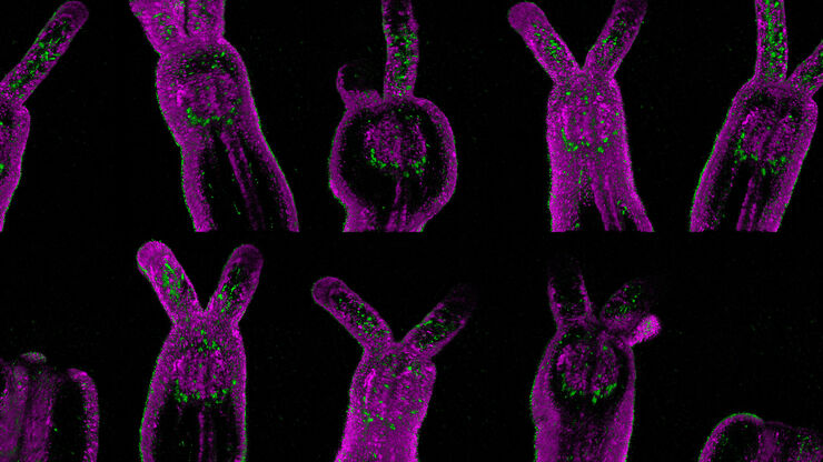
Nematostella

Live Cell Imaging Gallery

Live cell microscopy techniques are fundamental to get a better understanding of cellular and molecular function. Today, widefield microscopy is the most common technique used to visualize cell…

Tissue Image Gallery

Visual analysis of animal and human tissues is critical to understand complex diseases such as cancer or neurodegeneration. From basic immunohistochemistry to intravital imaging, confocal microscopy…

Super-Resolution Microscopy Image Gallery

Due to the diffraction limit of light, traditional confocal microscopy cannot resolve structures below ~240 nm. Super-resolution microscopy techniques, such as STED, PALM or STORM or some…
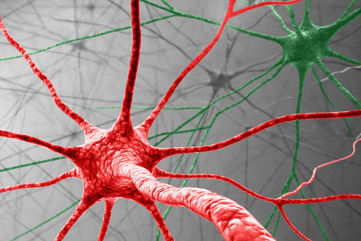
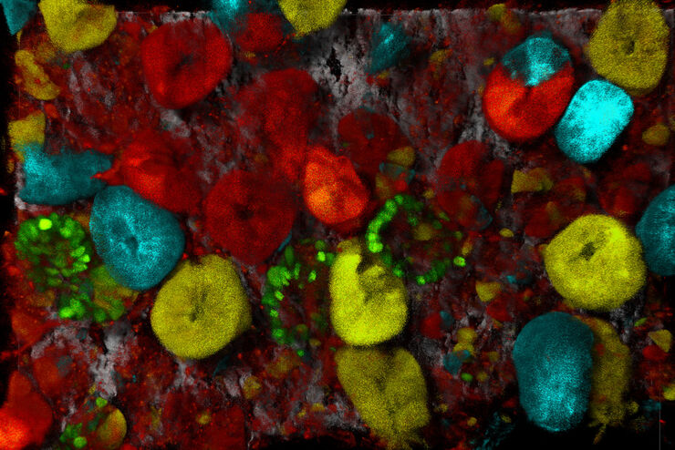
Virally labeled neurons (red) and astrocytes (green) in a cortical spheroid derived from human induced pluripotent stem cells. THUNDER Model Organism Imagerwith a 2x 0.15 NA objective at 3.4x zoomwas used to produce this 425 μm Z-stack (26 positions), which is presented here as an Extended Depth of Field(EDoF)projection.

Neuroscience Images

Neuroscience commonly uses microscopy to study the nervous system’s function and understand neurodegenerative diseases.

Multicolor Image Gallery

Fluorescence multicolor microscopy, which is one aspect of multiplex imaging, allows for the observation and analysis of multiple elements within the same sample – each tagged with a different…

Cancer Research Image Gallery

Fluorescence microscopy allows the study of changes occurring in tissue and cells during cancer development and progression. Techniques such as live cell imaging are critical to understand cancer…

Cell Biology Image Gallery

Cell biology studies the structure, function and behavior of cells, including cell metabolism, cell cycle, and cell signaling. Fluorescence microscopes are an integral part of a cell biologist…

Dissecting Proteomic Heterogeneity of the Tumor Microenvironment

This lecture will highlight cutting edge applications in applying laser microdissection and microscaled quantitative proteomics and phosphoproteomics to uncover exquisite intra- and inter-tumor…
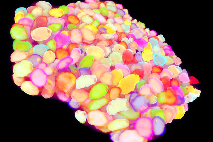
Pollen Flower - Taken with a 20x/0.8 objective, area of 6mm² with a depth of 100μm. 15 stitched tiles with 4 colors (DAPI/GFP/TRITC/Cy5) - a total of 13020 images. Video courtesy of James Marr, Leica Microsystems, USA

Developmental Biology Image Gallery

Developmental biology explores the development of complex organisms from the embryo to adulthood to understand in detail the origins of disease. This category of the gallery shows images about…

Kinetochore Assembly during Mitosis with TauSTED on 3D

Three-dimensional organization of the mitotic spindle together with the distribution of CENP-C and BUB1 based on TauSTED with multiple STED lines (592, 660 and 775 nm) can provide insights…

How to Quantify Changes in the Metabolic Status of Single Cells

Metabolic imaging based on fluorescence lifetime provides insights into the metabolic dynamics of cells, but its use has been limited as expertise in advanced microscopy techniques was needed. Now,…

Putting Dynamic Live Cell Data into the Ultrastructural Context

With workflow Coral Life, searching for a needle in the haystack is a thing of the past. Take advantage of correlative light and electron microscopy to identify directly the right cell at the right…

AI in Microscopy Webinar

We demonstrate residual channel attention networks for restoring and enhancing volumetric time-lapse (4D) fluorescence microscopy data.

20 Years of Leica Laser Microdissection

Phenotype-genotype correlations are key for insight. From Eye to Insight is therefore fitting perfectly to Leica Microsystems and in particular to laser microdissection. Laser Microdissection, also…
Virally labeled neurons (red) and astrocytes (green) in a cortical spheroid derived from human induced pluripotent stem cells. THUNDER Model Organism Imager with a 2x 0.15 NA objective at 3.4x zoom was used to produce this 425 µm Z-stack (26 positions), which is presented here as an Extended Depth of Field (EDoF) projection.  Images courtesy of Dr. Fikri Birey  from the Dr. Sergiu Pasca laboratory at Stanford University, 3165 Porter Dr., Palo Alto, CA

Download The Guide to Live Cell Imaging

In life science research, live cell imaging is an indispensable tool to visualize cells in a state as in vivo as possible. This E-book reviews a wide range of important considerations to take to…
3D reconstruction of an intercellular bridge in a C. elegans embryo

Download EM Workflow Solutions Booklet

This publication is a compilation of appropriate workflows for the most frequently used sample preparation methods, like Correlative Methodologies, Optogenetics & Electro-Physiology, Surface Analysis,…

Exploring the Structure and Life Cycle of Viruses

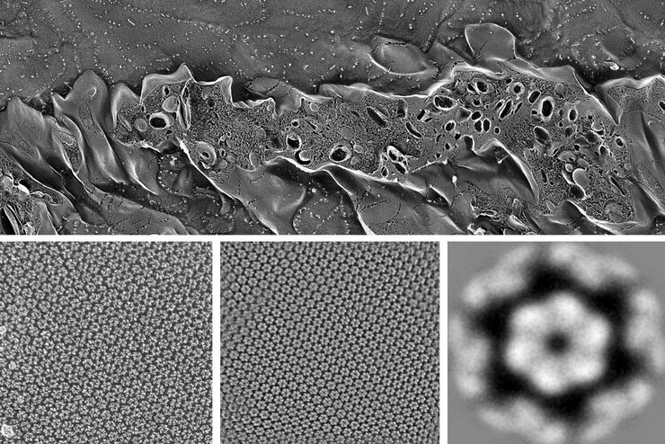
The SARS-CoV-2 outbreak started in late December 2019 and has since reached a global pandemic, leading to a worldwide battle against COVID-19. The ever-evolving electron microscopy methods offer a…
Product image EM TIC 3X

Application Booklet for EM TIC 3X

Today, ion beam milling is one of the most widely-used methods for preparing samples for electron microscopy. Download this 76-pages booklet today and learn how to improve your processes.

Regulators of Actin Cytoskeletal Regulation and Cell Migration in Human NK Cells

Dr. Mace will describe new advances in our understanding of the regulation of human NK cell actin cytoskeletal remodeling in cell migration and immune synapse formation derived from confocal and…

Adding Dimensions to Multiplex Molecular Imaging

Molecular imaging of living specimens offers a means to draw upon the growing body of high-throughput molecular data to better understand the underlying cellular and molecular mechanisms of complex…

Understanding Motor Sequence Generation Across Spatiotemporal Scales

We have developed a microscopy-based pipeline to characterize a developmentally critical behavior at the pupal stage of development, called the ecdysis sequence. We study brain-wide neuronal activity…
Root-hypocotyl junction of Arabidopsis thaliana. Image acquired with TauContrast. Sample courtesy: Dr. Melanie Krebs, COS, University of Heidelberg.

Benefits of TauContrast to Image Complex Samples

In this interview, Dr. Timo Zimmermann talks about his experience with the application of TauSense tools and their potential for the investigation of demanding samples such as thick samples or…
Scroll to top