Leica Microsystems

Leica Microsystems

Leica Microsystems is a world leader in microscopes and scientific instruments. Founded as a family business in the nineteenth century, the company’s history was marked by unparalleled innovation on its way to becoming a global enterprise.

Its historically close cooperation with the scientific community is the key to Leica Microsystems’ tradition of innovation, which draws on users’ ideas and creates solutions tailored to their requirements. At the global level, Leica Microsystems is organized in three divisions, all of which are among the leaders in their respective fields: Life Science, Industry and Medical.

The company is represented in over 100 countries with 6 manufacturing facilities in 5 countries, sales and service organizations in 20 countries, and an international network of dealers. The company is headquartered in Wetzlar, Germany.
http://www.leica-microsystems.com/

Why is Manual Visual Inspection of Medical Devices so Challenging?

This article discusses how manual visual inspection, which is prevalent in the medical device industry, can lead to inconsistent results. It also addresses the challenges quality managers and…

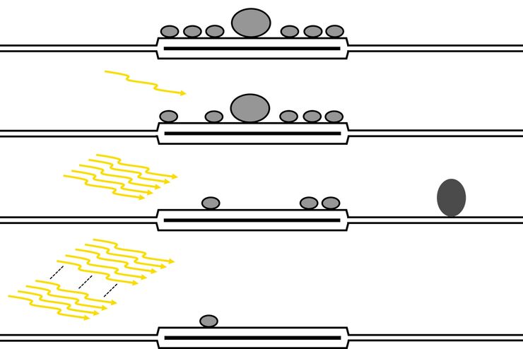
Fast, High-quality Vitrification with the EM ICE High Pressure Freezer

The EM ICE High Pressure Freezer was developed with a unique freezing principle and uses only a single pressurization and cooling liquid: liquified nitrogen (LN2). This design enables three major…
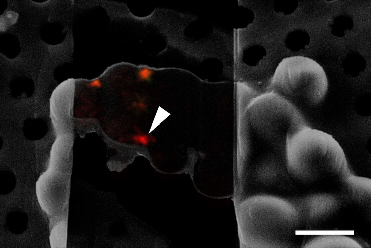
Cryo FIB lamella - Overlay of SEM and confocal fluorescence image. Target structure in yeast cells (nuclear pore proteine Nup159-Atg8-split Venus, red) marked by an arrow. Scale bar: 5 µm. Alegretti et al.,  Nature 586, 796-800 (2020).

Targeting Active Recycling Nuclear Pore Complexes using Cryo Confocal Microscopy

In this article, how cryo light microscopy and, in particular cryo confocal microscopy, is used to improve the reliability of cryo EM workflows is described. The quality of the EM grids and samples is…

Advanced Visualization in ENT Surgery

ENT surgery poses unique challenges. It requires excellent visualization, both in terms of magnification and illumination. As such, choosing the right operating microscope is critical.

Advanced Visualization in Spine Surgery

There have been meaningful advances in spine surgery in recent years. Minimally Invasive Spine Surgery (MISS) techniques provide significant benefits, helping to minimize surgical trauma, improve…

How Augmented Reality is Transforming Vascular Neurosurgery

Augmented Reality is changing surgery, with new information helping to improve the precision and safety of procedures. This is especially true in vascular neurosurgery where Augmented Reality is…

Overcoming Ophthalmologic Surgery Challenges

Ophthalmology surgical procedures involving both the anterior and posterior segment can be particularly challenging. Good visualization is required to operate with precision and confidence. Prof.…

Advanced Visualization in Maxillofacial Plastic and Reconstructive Surgery

Plastic and Reconstructive Surgery is very demanding. Surgical microscopes play an important role, helping to ensure flaps are well vascularized. Dr. Christine Bach is a Plastic and Reconstructive…
Dividing fission yeast S. pombe stained with two markers against spindle pole bodies (Pcp1-GFP, green) and cytokinesis ring (Rlc1-mCherry; red).

Studying Cell Division

Cell division is a biological process during which all cellular components must be distributed among the daughter cells. The division process requires firm coordination for success. Microscopy is…

Investigating Synapses in Brain Slices with Enhanced Functional Electron Microscopy

A fundamental question of neuroscience is: what is the relationship between structural and functional properties of synapses? Over the last few decades, electrophysiology has shed light on synaptic…

GLOW800 Augmented Reality Fluorescence in AVM (Arteriovenous Malformation) Treatment

In this case study Prof. Dr. Feres Chaddad talks about the treatment of AVMs. It illustrates how the Augmented Reality Fluorescence GLOW800 can help surgeons during microsurgical resection by…
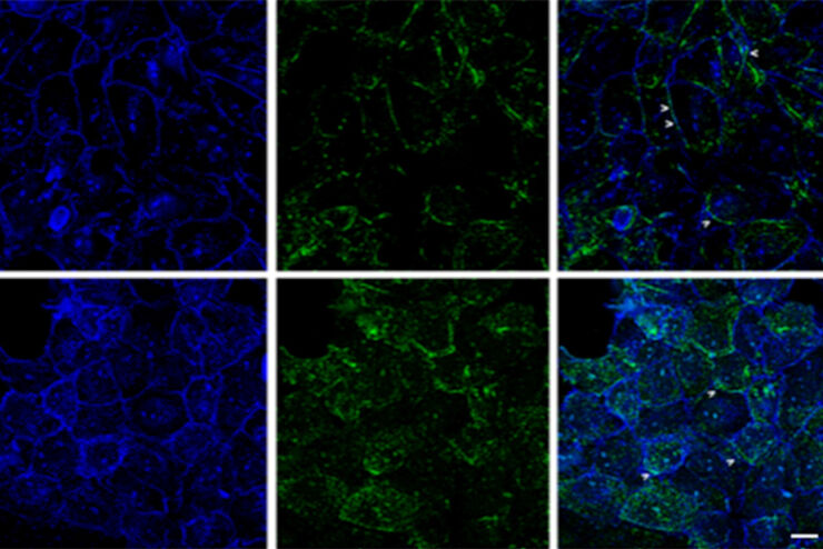
Mouse kidney section with Alexa Fluor™ 488 WGA, Alexa Fluor™ 568 Phalloidin, and DAPI. Sample is a FluoCells™ prepared slide #3 from Thermo Fisher Scientific, Waltham, MA, USA. Images courtesy of Dr. Reyna Martinez – De Luna, Upstate Medical University, Department of Ophthalmology.

The Power of Pairing Adaptive Deconvolution with Computational Clearing

Learn how deconvolution allows you to overcome losses in image resolution and contrast in widefield fluorescence microscopy due to the wave nature of light and the diffraction of light by optical…

Improvement of Imaging Techniques to Understand Organelle Membrane Cell Dynamics

Understanding cell functions in normal and tumorous tissue is a key factor in advancing research of potential treatment strategies and understanding why some treatments might fail. Single-cell…

Neurovascular Surgery & Augmented Reality Fluorescence

Vascular neurosurgery is highly complex. Surgeons need to be able to rely on robust anatomical information. As such, visualization technologies play an essential role. Prof. Nils Ole Schmidt is a…
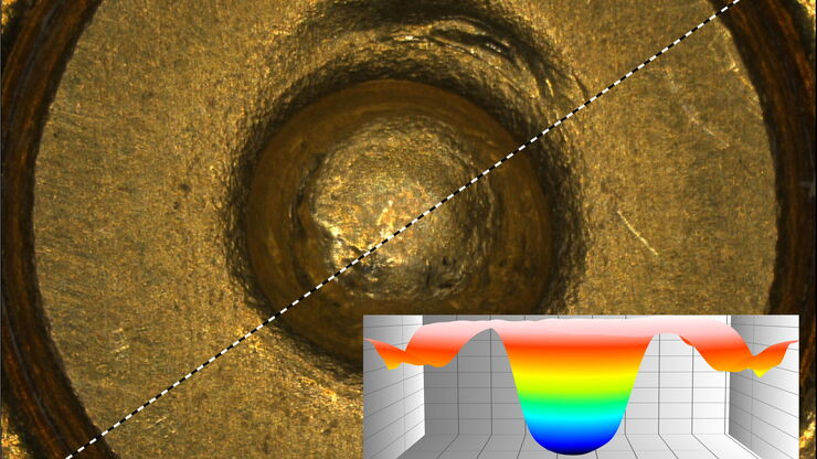
Topographic analysis of firing pin.

Topographic Analysis of Firing Pin Impressions on Cartridge Cases

The analysis of fired cartridges for primer cup morphology and flattening and firing pin impression (crater) depth using topographic data is discussed in this article. Topographical analysis of the…
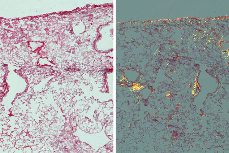
Mouse lung sections

Studying Pulmonary Fibrosis

The results shown in this article demonstrate that fibrotic and non-fibrotic regions of collagen present in mouse lung tissue can be distinguished better with polarized light compared to brightfield.…

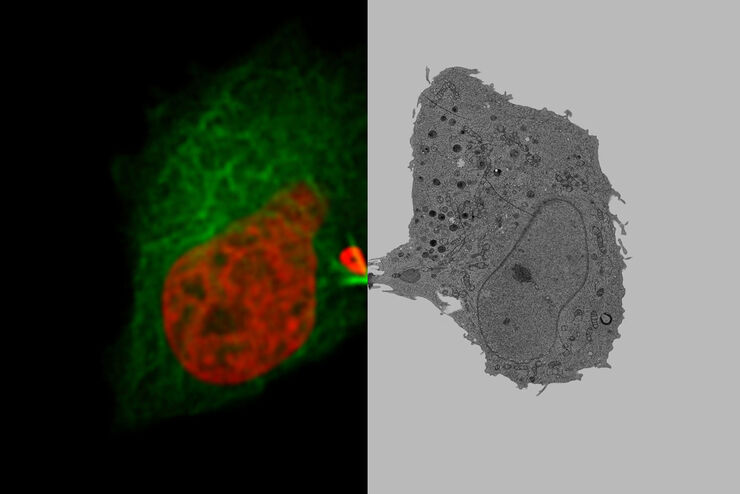
Advancing Cell Biology with Cryo-Correlative Microscopy

Correlative light and electron microscopy (CLEM) advances biological discoveries by merging different microscopes and imaging modalities to study systems in 4D. Combining fluorescence microscopy with…
Mouse lymphnode acquired with a THUNDER Imager 3D Cell Culture. Image courtesy of Dr. Selina Keppler, Munich, Germany.

Image Gallery: THUNDER Imager

To help you answer important scientific questions, THUNDER Imagers eliminate the out-of-focus blur that clouds the view of thick samples when using camera-based fluorescence microscopes. They achieve…

Keeping Particulate Contamination Under Control in Pharmaceutical Products

This article describes how a 2-methods-in-1 solution combining optical microscopy and laser induced breakdown spectroscopy (LIBS) can be utilized for identification of particulate contaminants in the…

Fast Visual and Chemical Analysis of Contamination and Underlying Layers

Visual and chemical analysis of contamination on materials with a 2-methods-in-1 solution leading to an efficient, more complete analysis workflow is described in this report. A 2-in-1 solution,…

Hand-Held OCT Clinical & Research Applications

Hand-held Optical Coherence Tomography (OCT) has revolutionized pediatric ophthalmology and has had a significant impact on ophthalmology in general. The Leica Envisu C2300 hand-held OCT allows to…

From Organs to Tissues to Cells: Analyzing 3D Specimens with Widefield Microscopy

Obtaining high-quality data and images from thick 3D samples is challenging using traditional widefield microscopy because of the contribution of out-of-focus light. In this webinar, Falco Krüger…

How does an Automated Rating Solution for Steel Inclusions Work?

The rating of non-metallic inclusions (NMIs) to determine steel quality is critical for many industrial applications. For an efficient and cost-effective steel quality evaluation, an automated NMI…

Perform Microscopy Analysis for Pathology Ergonomically and Efficiently

The main performance features of a microscope which are critical for rapid, ergonomic, and precise microscopic analysis of pathology specimens are described in this article. Microscopic analysis of…

How to improve your Biomarker Discovery Workflow with Laser Microdissection

Biomarkers can be used as indicators of certain diseases, such as cancer. The tumor microenvironment moved into the spotlight in this concern. It is in close interaction with the tumor itself.…

How to Conduct Standard-Compliant Analysis of Non-Metallic Inclusions in Steel

This webinar will provide an overview of the significance of non-metallic inclusions in steel and outline the important global standards for rating the quality of steel and difficulties that arise in…
Oncological Maxillofacial Reconstructive Surgery at the Paris Saint-Joseph Hospital with Dr. Benichou. The Leica M530 OHX microscope is used by plastic surgeons in a comfortable standing up position.

Plastic & Reconstructive Surgery: Why Use a Microscope

Plastic and Reconstructive Surgery procedures can be delicate. Visualization solutions play an essential role, allowing to perform the surgery in the best conditions. And more and more plastic…

Minimally Invasive Spine Surgery: Improving Precision and Accuracy with Microscopes

Spine surgery is extremely delicate and requires extensive training and experience. Innovative visualization technologies can also help achieve better outcomes allowing to see more and have a clearer…

Neurosurgery with Heads-up Display

In the following video interviews Prof. Dr. Raphael Guzman, Vice Chairman of the Department of Neurosurgery at the University Hospital in Basel, Switzerland, talks about his experience in heads-up…

Workflows and Instrumentation for Cryo-electron Microscopy

Cryo-electron microscopy is an increasingly popular modality to study the structures of macromolecular complexes and has enabled numerous new insights in cell biology. In recent years, cryo-electron…
Scroll to top