
Life Science Research
Life Science Research
This is the place to expand your knowledge, research capabilities, and practical applications of microscopy in various scientific fields. Learn how to achieve precise visualization, image interpretation, and research advancements. Find insightful information on advanced microscopy, imaging techniques, sample preparation, and image analysis. Topics covered include cell biology, neuroscience, and cancer research with a focus on cutting-edge applications and innovations.
Filter articles
Tags
Story Type
Products
Loading...
How Does The Cytoskeleton Transport Molecules?
VIDEO ON DEMAND - See how 3D cysts derived from MDCK cells help scientists understand how proteins are transported and recycled in tissues and the role of the cytoskeleton in this transport.
Loading...
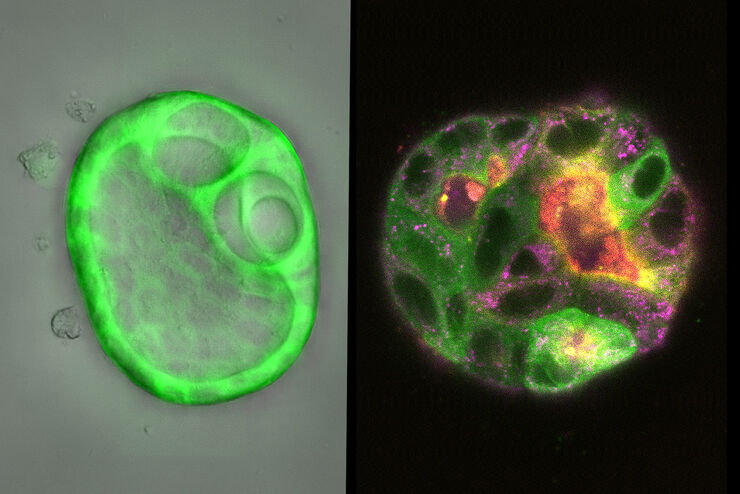
 embryo, from sphere stage to somite stages.")
Studying Early Phase Development of Zebrafish Embryos
This video on demand focuses on combining widefield and confocal imaging to study the early-stage development of zebrafish embryos (Danio rerio), from oocyte to multicellular stage.
Loading...

Multi-Color Caspase 3/7 Assays with Mica
Caspases are involved in apoptosis and can be utilized to determine if cells are undergoing this programmed cell death pathway in so-called caspase assays. These assays can be run by e.g. flow…
Loading...

How To Get Multi Label Experiment Data With Full Spatiotemporal Correlation
This video on demand focuses on the special challenges of live cell experiments. Our hosts Lynne Turnbull and Oliver Schlicker use the example of studying the mitochondrial activity of live cells.…
Loading...

Find Relevant Specimen Details from Overviews
Switch from searching image by image to seeing the full overview of samples quickly and identifying the important specimen details instantly with confocal microscopy. Use that knowledge to set up…
Loading...

AI Microscopy Image Analysis – An Introduction
Artificial intelligence-guided microscopy image analysis and visualization is a powerful tool for data-driven scientific discovery. AI can help researchers tackle challenging imaging applications,…
Loading...

How to Target Fluorescent Structures in 3D for Cryo-FIB Milling
This article describes the major steps of the cryo-electron tomography workflow including super-resolution cryo-confocal microscopy. We describe how subcellular structures can be precisely located in…
Loading...

Precise 3D Targeting for EM Imaging - Access What Matters
Find out how the seamless cryo-electron tomography workflow Coral Cryo uses confocal super resolution to target your structure of interest more precisely.
Loading...
, unsaturated lipids (magenta, 3050 cm-1), collagen (SHG, cyan). Sample courtesy of R. Rudolf, J Klicks, Hochschule Mannheim")
The Potential of Coherent Raman Scattering Microscopy at a Glance
Coherent Raman scattering microscopy (CRS) is a powerful approach for label-free, chemically specific imaging. It is based on the characteristic intrinsic vibrational contrast of molecules in the…
