A Guide to Model Organisms in Research

Go from overview to fine detail to enhance the understanding of your model organisms with flexible imaging systems from Leica Microsystems.

A model organism is a species used by researchers to study specific biological processes. They have similar genetic characteristics to humans and are commonly used in research areas such as genetics, developmental biology, and neuroscience.  Model organisms are typically chosen for their easy maintenance and reproduction in a laboratory setting, short generation cycles, or the capacity to generate mutants to study certain traits or diseases.

Model organisms provide valuable insights into biological systems at the cell, tissue, organ, and system level. There are several types of model organisms and they vary in complexity and use. Small, simple organisms, such as yeast, are typically used to study gene mutations in human cancers, whereas the fruit fly (Drosophila melanogaster) and the zebrafish (Danio rerio) are ideal for the study of genetics and development of disease. Mouse models are also extensively used in biomedical research to study the progress of disease and development of new drugs.

Model Organism Imaging

Go from overview to fine detail to enhance the understanding of your model organisms with flexible imaging systems from Leica Microsystems.

Leica Microsystems provides innovative microscopy solutions to achieve optimal image quality. These solutions are applicable even with large and complex specimens, such as model organisms, and include platforms for screening and manipulation.
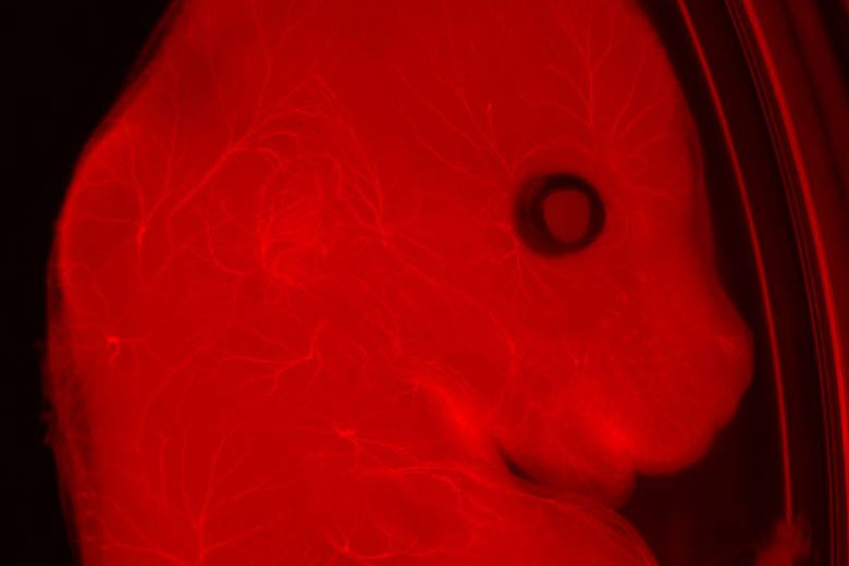
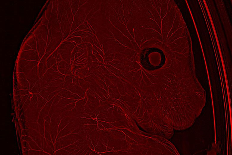
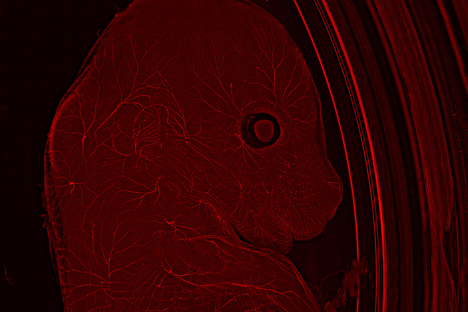
Mouse embryo Mouse embryo with THUNDER Imager Model Organism

Neurofilaments stained in red to assess neuronal outgrowth in E12-14 mouse. The mouse was uncleared. Courtesy of Yves Lutz, Centre d’imagerie, IGBMC, France.

Microscopy for Model Organism Research

Model organisms are in most cases large, complex, and dense, which can be a challenge. Therefore, there is a need for flexibility from microscope systems to enable the study of these samples. Traditionally, several microscopy techniques are necessary for the study of model organisms, from stereomicroscopes that enable macroscopic analysis and manipulation of specimens such as zebrafish, to multiphoton microscopes that allow deep imaging of large live organisms.

For these thicker specimens, confocal microscopy is the tool of choice to see detail at depth and avoid out-focus-signals. However, it usually poses several challenges, such as slow speeds and photobleaching due to high laser power.

Innovative techniques, such as light sheet microscopy, are becoming more prominent when imaging these light-sensitive samples in 3D thanks to its low phototoxicity.

Challenges When Imaging Model Organisms

Work involving model organisms is very heterogeneous and, as such, challenges when using microscopy techniques vary depending on whether they are used for dynamic studies or the study of molecular events, but it can also depend on the complexity of the model organism used, going from the few cells of C. elegans to large complex animals such as the mouse.

A common challenge encountered when imaging model organisms is speed of throughput. Due to the size of the sample, imaging can involve lengthy acquisition times and the analysis of many images to get a complete dataset, which overall reduces experimental efficiency. Speed of acquisition is also an issue when imaging live model organisms, as phototoxicity and photodamage can result in disruption of the sample’s physiology or even cell or sample death. Finally, imaging points within the deep layers of the sample can also produce image artifacts and blurring.
Related articles

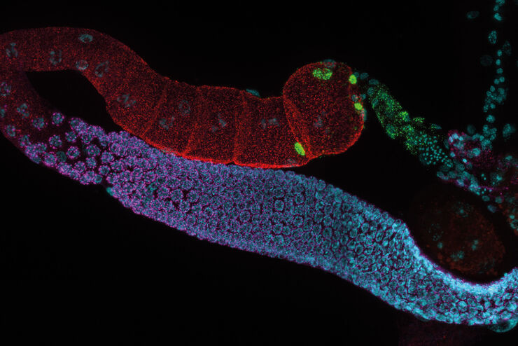
C. elegans Gonades - THUNDER Imager  Adult hermaphrodit, Staining: blue - DAPI (Nucleus), green - SP56 (sperms), red - RME-2 (oocyte), mangenta - PGL-1 (RNA + protein granules) Image courtesy of Prof. Dr. Christian Eckmann, Martin Luther University, Halle, Germany

Physiology Image Gallery

Physiology is about the processes and functions within a living organism. Research in physiology focuses on the activities and functions of an organism’s organs, tissues, or cells, including the…
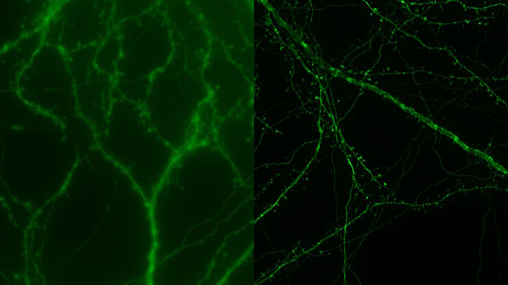
Mouse cortical neurons. Transgenic GFP (green). Image courtesy of Prof. Hui Guo, School of Life Sciences, Central South University, China

How Microscopy Helps the Study of Mechanoceptive and Synaptic Pathways

In this podcast, Dr Langenhan explains how microscopy helps his team to study mechanoceptive and synaptic pathways, their challenges, and how they overcome them.
Brain organoid section (DAPI) acquired using THUNDER Imager Live Cell. Image courtesy of Janina Kaspar and Irene Santisteban, Schäfer Lab, TUM.

Imaging Organoid Models to Investigate Brain Health

Imaging human brain organoid models to study the phenotypes of specialized brain cells called microglia, and the potential applications of these organoid models in health and disease.

THUNDER Imagers: High Performance, Versatility and Ease-of-Use for your Everyday Imaging Workflows

This webinar will showcase the versatility and performance of THUNDER Imagers in many different life science applications: from counting nuclei in retina sections and RNA molecules in cancer tissue…
Scroll to top