The Experiment
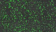
In this video, our expert image analyst Luciano Lucas illustrates how to use Mica’s AI-enabled software to start the 3D image analysis process. Using a 3D dataset of the unicellular organism Paremecium tetaurella, we learn how to segment 4 different structures including the cilia, nucleus and basal bodies. We then explore the spatial relationships between the different visible biological elements and learn how to create graphs and charts quantifying these relationships. Luciano demonstrates different options for presenting data including colour coding that allows you to best communicate your findings to your audience. The session concludes with the creation of high-fidelity video animations and other publication ready outputs.


