
How Mica benefits the research of Asherman’s syndrome
Asherman’s syndrome is a dreadful disorder of the uterus which can lead to infertility. Despite extensive research, reproducible treatments to improve the structural and functional morphology of the affected endometrium have not been established.
Professor Kang’s group is working to verify the efficacy of transplanting organoids into murine disease models by investigating the underlying molecular mechanisms. They recently published a paper in the journal Theranostics. It focuses on how endometrial organoids serve as a reservoir of functional mitochondria for uterine repair.
Their research findings suggest that endometrial organoid-originating mitochondria might be key players to mediate uterine repair resulting in fertility enhancement by recovering abrogated metabolic circumstance of endometrium with the disorder.
Since the publication of the article in Theranostics, we have conducted 9 interviews with Prof. Kang regarding how the use of the Mica Microhub has benefited her research group. The Mica Microhub has generally made microscopy more convenient for her team. For example, it significantly reduces microscope usage time by enabling high-speed data acquisition. Its ease of use allows effectively anyone to operate it. Additionally, it makes it easy to focus on tissue samples with uneven thickness during imaging. Another notable feature is its ability to automatically perform water immersion with high-magnification objective lenses when needed.
Read the interview with Professor Youn-Jung Kang
Prof. Kang, please tell us about your research group with a simple overview and background.
Prof. Kang: Our research group investigates crucial factors that determine the uterine microenvironment (called uterine receptivity), wherein the embryo implants and pregnancy is maintained successfully. We have been working on developing novel therapeutic strategies to regenerate the endometrial functionalis for patients suffering from repeated implantation failure induced by various endometrial diseases. To address one of our interests, we have applied 3D endometrial organoids and defined the cellular and molecular mechanisms underlying their remarkable regenerative capacity in the endometrium with Asherman's Syndrome.
Could you first tell us about the primary focus of your research and why microscopy is important?
Prof. Kang: Because our study is focused on evaluating the efficacy of transplanted organoids by histological analyses, a device capable of imaging high-resolution shots and efficiently photographing a large number of data by serial magnifications was essentially required. The equipment that met our demand was Mica.
What application or experiment are you trying to do using Mica?
Prof. Kang: The uteri are horizontally long like an intestine, thus we had a difficulty to observe them as a whole using our conventional microscope. We had to take several dozen images and pile them one by one manually, adjusting the microscope directly to observe its overall appearance. However, Mica takes a whole section in one shot and high-resolution images were obtained in a very short imaging time. Mica shortened our analysis time and enabled us to conduct further analyses through the whole section.
What impact does it have if you do not successfully accomplish these applications or experiments?
Prof. Kang: By coincidence, we were able to use a Mica system during the revision period prior to publication. In the process of analyzing additional data, we were able to obtain sufficient data due to the fast speed of Mica and finally finish the revision within the deadline. If we had just used our conventional microscope, we would have not been able to meet the deadline.
Did Mica help you solve the problem? If so, how?
Prof. Kang: Yes, using Mica we took lots of high-resolution images of whole sections with high-speed. In addition, it was very easy to utilize and perform all the analyses, because it was equipped with a simple software which was easy to use.
What do you like the most about Mica?
Prof. Kang: Unlike other microscopes, it was very convenient because the work of the user after loading the sample was relatively small. For example, when shooting at a high magnification, immersion water was automatically sprayed, resulting in little user inconvenience.
By solving these problems, what value and benefit were you able to gain?
Prof. Kang: When histological staining was performed, the thickness of the sections may not be constant. In this case, the focus may vary in one slide, resulting in many difficulties with imaging. However, Mica provided the average focus after random measurements. In addition, if spraying oil and water was done directly by users, manual operation might be inevitable due to a frequent shifting of the focus and position. Mica solved this inconvenience, saving us time.
What are other possible applications you can envision doing with Mica?
Prof. Kang: We are conducting a follow-up study on endometrial organoid dynamics in the process of regeneration. We think it will be helpful for our future research if we were able to observe real-time morphological changes using the live-cell imaging function.
To finish, in a couple of sentences, how would you summarize your research results?
Prof. Kang: Asherman's syndrome (AS) is a dreadful gynecological disorder of the uterus characterized by intrauterine adhesion with severe fibrosis. Despite extensive research on overcoming AS, evidence-based effective treatments have not been established. So, we successfully generated human/mouse endometrial organoids and transplanted them into mouse AS models. Transplanted organoids showed anti-fibrosis and an alleviated endometrial environment, resulting in recovery of fertility. In addition, we uncovered that endometrial organoid-originating mitochondria might be key players to mediate the uterine repair by recovering abrogated metabolic circumstance.
Read the research paper:
Endometrial organoids: a reservoir of functional mitochondria for uterine repair
Theranostics 2024; 14(3):954-972. doi:10.7150/thno.90538

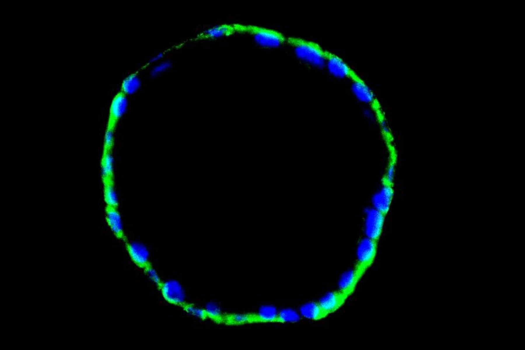
, tubulin with Cy5 (red), and nuclei with DAPI (blue). Image courtesy of Dr. Günter Giese, Max Planck Institute for Medical Research, Heidelberg, Germany.")

