Traditional versus AI confluency assessment methods
There are significant limitations when assessing confluency via manual cell-culture inspection and image analysis. This disadvantage is especially true for cases with complex cell morphologies, crowded cells, and dynamic experimental conditions. Manual assessment is prone to human subjectivity and error. So there is a serious risk of inconsistent results.
This significant limitation can hinder the precision and reliability of confluency measurements. Hence there is a need for advanced solutions which use AI technologies like automated image analysis and deep learning to make cell confluency assessment efficient and reproducible, especially when dealing with dynamic and diverse cellular environments.
Advantages of confluency measurement with AI
AI-based confluency assessment offers the following advantages:
- Adaptability to diverse cell morphologies thanks to advanced models like convolutional neural networks (CNNs)
- Experimental flexibility when working with complex and diverse cell lines
- Dynamic response to changing experimental conditions enabling researchers to have a more nuanced understanding of confluency in dynamic cellular environments
- Edge detection for complex and crowded cultures allowing cell boundaries to be clearly identified
- Robust and reliable analysis which outperforms manual methods
Additional benefits with AI
- Determine confluency efficiently and save valuable time compared to when using manual methods
- Accurate confluency analysis even for unique or complex cell lines
- Increased reproducibility and consistent results thanks to standardized confluency measurements
- Optimized experimental conditions for cell culture with real-time feedback during confluency analysis
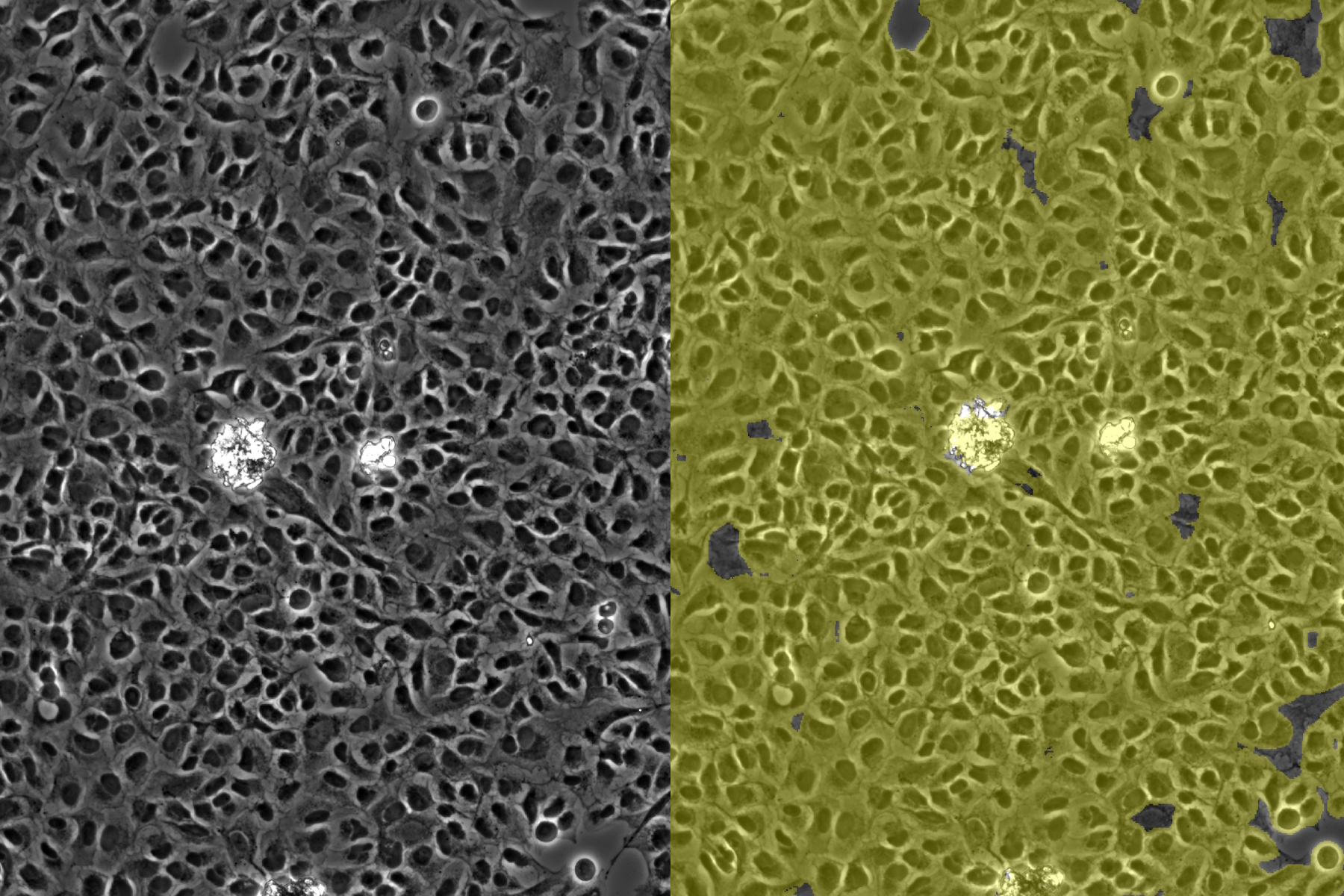
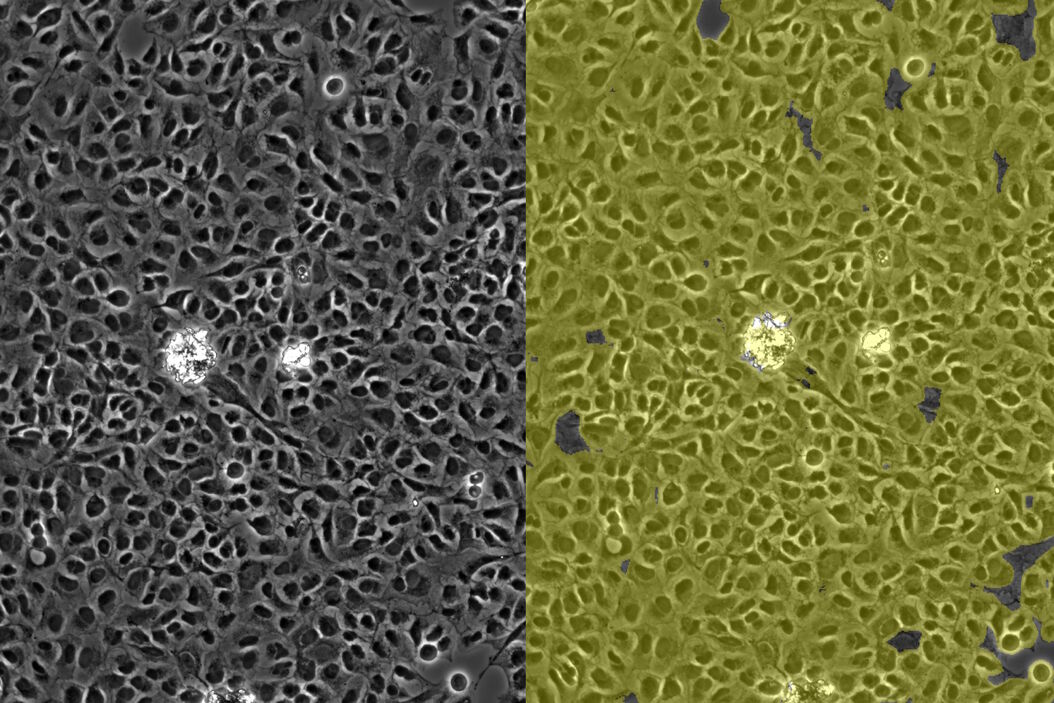
Both manual and AI-based cell confluency measurements performed with the Mateo FL microscope are shown below. The confluency values are from manual assessment of conventional phase-contrast images of cell culture (left) and AI-assisted analysis (right).


