Probing vibrational motions of molecules
The chemical bonds in molecules can shake, bend and rattle. They make these motions at particular rates or frequencies. These frequencies are so particular that we can identify what kind of chemical bond is rattling by tuning into their particular vibrational frequencies. For instance, many organic molecules contain bonds between carbon and hydrogen atoms, and thus they have C-H vibrational motions. More importantly, the C–H vibrational frequency is very different from the oscillatory motions of other chemical bonds, such as O–H, the oxygen-hydrogen bond. In other words, by examining the vibrational frequencies of a molecule, we can say something significant about chemical structure of the molecule. A vibrational analysis thus corresponds to a chemical analysis.
Some of these vibrational motions can be addressed by examining molecules with light. Unfortunately, the frequencies of molecular vibrations are much lower than the oscillation frequencies of visible light waves. This means that we cannot directly tune into these molecular motions with common light sources, including the type of lasers that are part of optical microscopes. However, the molecules can be inspected in an indirect way. Raman spectroscopy is an example of such an indirect inspection [1]. In Raman spectroscopy and microscopy, a conventional laser beam addresses the sample and the light that is scattered off the molecules is analyzed. The scattered light that is of a different color than the excitation light may contain information about the molecular vibration. This approach looks very similar to a fluorescence measurement, but the information collected is very different. In Raman spectroscopy, the difference between the frequency of the laser light and the Raman scattered light corresponds to the frequency of the chemical bonds. Using appropriate filters and spectrometers, it is possible to confidently collect the Raman-scattered light in an optical microscope, and thus gather chemical information about the sample. This offers unique possibilities. Since virtually every molecule exhibits particular chemical bond vibrations that are Raman active, it is possible to generate chemical maps of samples without the need of any extrinsic labels. Not surprisingly, this capability has sparked the interest of many research disciplines, including material synthesis, forensic research, mineralogy, toxicology, and the chemical analysis of artwork [2].
Raman spectroscopy and microscopy has also had an impact in biology. Raman microscopy enables the study of cells and tissues in a label-free manner, which opens opportunities towards studying biological samples that are difficult to stain or prepare for standard optical inspection. For instance, Raman spectroscopy has been shown to be sensitive enough to pick up subtle differences in the chemical makeup of healthy and cancerous tissues. Despite its enormous potential for biological research, Raman microscopy has not fully matured into a routine imaging technique in the biology laboratory. The reason for the limited impact of Raman microscopy in tissue and cell research is the intrinsic weakness of the Raman signal. The signal is weak because the Raman effect probes the molecules indirectly: the laser light is not in resonance with the molecular vibrations. The Raman effect is measurable, but, compared to fluorescence, it is a very weak effect. This implies that recording a Raman image takes much longer than taking a fluorescence image. Whereas most fluorescence images can be taken within a second, a Raman image of the same dimensions would require an hour or more. Clearly, such image acquisition times are not very attractive for biological imaging applications.
CARS microscopy enables new insights in biological and material samples
Does this mean that Raman microscopy has no future in the area of rapid biological imaging? Fortunately, there are alternative Raman techniques that significantly boost the signal levels. Nonlinear Raman techniques make use of ultrafast pulsed lasers, and probe the vibrational response of molecules in a more efficient way. Physically, nonlinear Raman techniques make the molecule vibrate in unison, generating coherent signals that can be up to five orders of magnitude higher compared to conventional Raman spectroscopy. There are several nonlinear Raman techniques, such as coherent anti-Stokes Raman scattering (CARS) and stimulated Raman scattering (SRS), all of which probe the same Raman-active molecular vibrations [3]. These techniques are by no means new: much of groundwork of the nonlinear Raman methods was laid in the 1960s. Within the family of nonlinear Raman methods, CARS is the first method that has grown into a user-friendly microscopy tool. The first CARS microscope dates back to 1982, but the first useful biological implementation was demonstrated a little over a decade ago, in 1999 [4]. Since then, the CARS microscopy technique has matured into an easy-to-use imaging tool, underlined by the release of the Leica TCS CARS microscope, which has added real-time vibrational contrast to the palette of the optical microscopist. The CARS imaging modality delivers images as fast and as crisp as what may be expected from a confocal fluorescence microscope. With such imaging capabilities at hand, a whole new domain of imaging applications is within reach.
Lipid and proteins
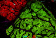
What is the range of applications of the CARS microscope? The CARS microscope has already made significant impact in the area of lipid imaging [5]. Lipids can be visualized by tuning into symmetric CH2 stretching vibration of aliphatic molecules. For examples, the CARS microscope can discriminate between saturated and unsaturated lipids, it can selectively detect cholesterol and cholesterol esters, and it can reveal information about the packing density of lipid membranes. The CARS microscope is sensitive enough to pick up signals from single phospholipids membranes, enabling the study of membrane biophysics, vesicle transport and organelle mapping. The strong CARS signal from lipids has also spurred research in lipid metabolism, trafficking of intracellular lipid bodies, and studies focused on the correlation between lipid accumulation and tumor growth. Most significantly, the strong CARS signal from lipid-rich myelin has revealed several important insights in the progression and treatment of neurodegenerate diseases.
Another vibrational response of interest is that of proteins. Although the CARS microscope is unable to conclusively discriminate among proteins, it is possible to generate maps of protein densities [6]. The CH3 methyl stretching vibration provides a convenient handle for mapping out protein distributions in tissues and cells. The spatial distribution of protein density is often and important marker in diseased tissue.
Diffusion of water and drugs
CARS microscopy has also been used as the preferred method of monitoring water dynamics in biological and synthetic systems [7]. The O–H stretching vibration provides a sensitive probe for water in tissues. This has enabled the study of water permeation in single cells and organelles, in addition to water dynamics in designed micro-structured materials.
The label-free and noninvasive character of CARS microscopy makes the technique ideally suited for monitoring permeation of topically applied chemicals through skin in vivo. CARS has been used to follow the diffusion of mineral oil through skin in animal models [8]; a method that has recently also been shown to work on skin of human patients [9].
Polymer films and nano-particles
Beyond the label-free chemical imaging of tissues and cells, the CARS microscope has been employed to visualize the chemical composition of polymer films and fabricated micro-structures. Contrast can be derived from the vibrational motions of the carbon network in polymers, or from carbonyl groups of organic molecules. The CARS signals from a many polymer structures are sufficiently strong to dial into a wide variety of vibrational Raman signatures for imaging purposes. In addition, the CARS microscope can probe molecular systems with strong absorptive features. Carbon nanotubes, for instance, can be visualized individually in the CARS microscope. Similarly, semiconducting nano-particles, such as silicon and iron oxide nano-structures can be visualized one-by-one based on their nonlinear CARS response [10]. Importantly, the CARS signals from these nano-particles are not subject to photobleaching, which guarantees sustained imaging of samples without fading of the contrast. These examples illustrate that there is a broad range of imaging applications for the CARS microscope in the material sciences.
Compared to the fluorescence microscope, the nonlinear Raman microscope is relatively young. Nonetheless, the impact of the nonlinear Raman technique can already be heard loud and clear, particularly in the area of label-free imaging of biological tissues. Rather than competing with fluorescence techniques, the ability to rapidly generate images based on vibrational contrast adds a new dimension to the existing contrast mechanisms of the optical microscope. The commercial availability of the CARS imaging modality gives the microscopist more options to examine the microscopic details of the illuminated sample. And with new contrast comes new discoveries. It is exciting to see how techniques like CARS microscopy can expand our understanding of the microworld.
References
- Krishnan KS and Raman CV: A new type of secondary radiation. Nature 121 (1928) 501–502.
- Turrell G and Corset J (eds): Raman Microscopy. Developments and Applications. Academic Press, San Diego (1996).
- Min W, Freudiger CW, Lu S and Xie XS: Coherent nonlinear optical microscopy: Beyond fluorescence microscopy. Annu. Rev. Phys. Chem. 62 (2011) 507–530.
- Zumbusch A, Holtom G and Xie XS: Vibrational Microscopy Using Coherent Anti-Stokes Raman Scattering. Phys. Rev. Lett. 82 (1999) 4142–4145.
- Le TT, Yue S and Cheng JX: Shedding new light on lipid biology with coherent anti-Stokes Raman scattering microscopy. J. Lipid Res. 51 (2010) 3091–3102.
- Benalcazar WA and Boppart SA: Nonlinear interferometric vibrational imaging for fas label-free visualization of molecular domains in skin. Anal. Bioanal. Chem. 400 (2011) 2817–2825.
- Potma EO, d Boeij WP, v Haastert PJM and Wiersma DA: Real-time vizualization of intracellular hydrodynamics. Proc. Natl. Acad. Sci. USA 98 (2001) 1577–1582.
- Evans CL, Potma EO, Puoris'haag M, Cote D, Lin C and Xie XS: Chemical imaging of tissue in vivo with video-rate coherent anti-Stokes Raman scattering (CARS) microscopy. Proc. Natl. Acad. Sci. USA 102 (2005) 16807–16812.
- Breunig HG, Bückle R, Kellner-Höfer M, Weinigel M, Lademann J, Sterry W and König K: Combined in vivo multiphoton and CARS imaging of healthy and disease-affected human skin. Microsc. Res. Techn., DOI: 10.1002/jemt.21082 (2011).
- Wang Y, Lin CY, Nikolaenko A, Raghunathan V and Potma EO: Four-wave mixing microscopy of nanostructures. Adv. Opt. Photon. 3 (2011) 1–52.


