About Live Cell Imaging
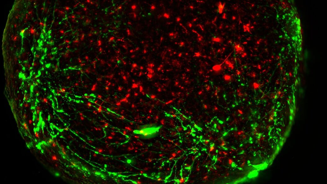
Besides the structural organization of cells or organs, dynamic processes are a major contributor to a functioning biological entity. Naturally, these processes can be best observed in living cells with non-invasive techniques like optical methods, collectively called “live-cell imaging” methods. Live-cell imaging covers all techniques where live cells are observed with microscopes – from the observation of embryogenesis with stereo microscopes, via cell growth studies with compound microscopes, until studies of physiological states of cells or cellular transport using fluorescent dyes or proteins. Although being highly demanding for both, experimenter and equipment (e.g. imaging systems, climate control), live-cell imaging techniques deliver results that are indispensable for present-day research.
Your Live Cell Imaging Needs
To perform successful live-cell imaging experiments, using the right platform is critical. When choosing an optical microscope for live‐cell imaging, the following 3 variables should be considered: detector sensitivity (signal‐to‐noise ratio), specimen viability, and image-acquisition speed.
Methods suitable for live-cell applications enable visualization of the dynamics without causing cell damage, as it can affect the results.
What’s inside
- What parameters need to be considered to maintain cell viability
- A full review of the wide range of Live Cell Imaging Techniques and Applications
- An insight into the problem of Out-of-Focus Light in Live Cell Microscopy and how to overcome it
- An introduction to the THUNDER Imagers and the DMi8 S platform that are ideally suited to cover all your live cell imaging applications.


