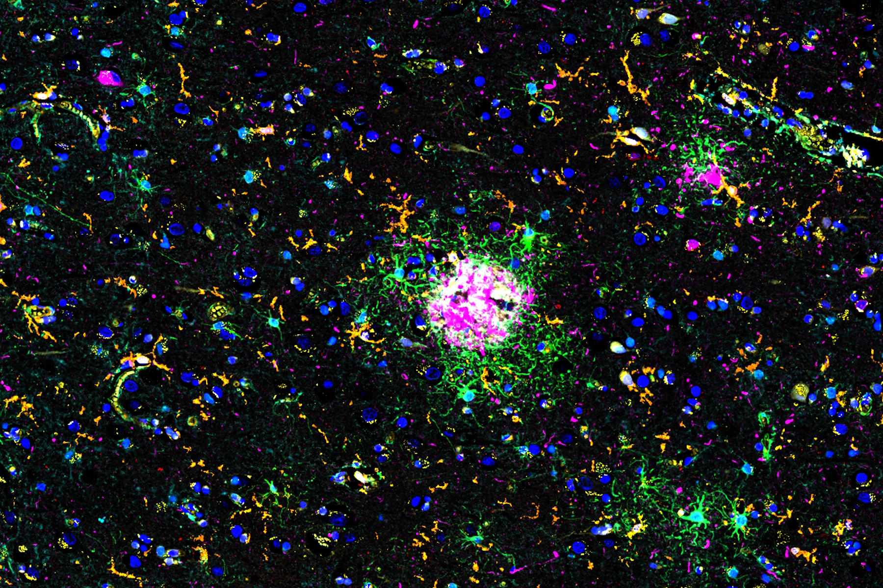
, microglia (TMEM119, IBA1), and Alzheimer’s-associated markers (β-amyloid and p-Tau217).")
Simplify demonstration and sharing of histopathology datasets
Alzheimer's disease, a genetic and sporadic neurodegenerative disorder, leads to cognitive decline in mid to late life and is pathologically characterized by β-amyloid plaques and phosphorylated tau tangles. With limited effective treatments available, novel investigative strategies are essential for progress in this field. By analyzing human brain tissue from Alzheimer's patients with the Cell DIVE multiplexed imaging solution, researchers can potentially uncover detailed histopathological features that may open new avenues for neuroscience research.
To highlight the capabilities of Cell DIVE, we provide several complete datasets accessible through the Minerva image viewer. Minerva is a lightweight, narrative-based image browser created by scientists at Harvard University to make demonstrating and sharing histopathology datasets easier for researchers and their colleagues [1]. Using Minerva, we have created a variety of guided narrations of different biological settings. This narration moves to different areas of interest in the various tissues and explains staining patterns and rationales for various biomarkers, outlining how a user might potentially build a Cell DIVE study and perform analysis. Using this application, users can assess the power of Cell DIVE imaging, brainstorm approaches to multiplexed imaging, and see how images might ultimately be transformed by analysis into quantitative data.
In this Minerva story, we identify several key findings. For example, we can examine the key pathophysiological feature of Alzheimer's disease, which is the presence of β-amyloid plaques and tangles of phosphorylated tau tangles. Using conjugated antibodies directed against these plaques and a cohort of tau proteins (p-Tau217, p-Tau181, Tau GT-83), we can observe the distribution of these structures throughout the tissue section. We observe that there is a higher concentration of tau and amyloid structures on the outer periphery of the brain section.
The Minerva image viewer reveals how Cell DIVE imaging translates into detailed data, providing researchers with an essential tool for innovating and refining multiplexed imaging methodologies.
See for yourself and explore the Alzheimer's interactive data set: