.")
Discover an end-to-end workflow for meaningful insights in advanced 3D cell cultures, whether you want to study organoids, spheroids, or stem cells.
The workflow presented starts with Mateo TL, which allows observation checks of organoid cell cultures easily and intuitively. The samples are then imaged using THUNDER Imager for a clear view of details deep within intact samples – in real time and without out-of-focus blur. This technology considers all relevant optical parameters, ensuring haze-free results. THUNDER Imagers excel in studying 3D cell cultures, capturing both static and dynamic cellular states with enhanced clarity.
For the study of 3D cell cultures, the end-to-end workflow showcases THUNDER Imagers' unique capabilities in automating assays to efficiently study next-generation disease models. The system allows imaging of large sample volumes, such as organoids, at high speed and with high sensitivity – ensuring low phototoxicity and photobleaching – for higher throughput under optimal conditions. Automation minimizes user interaction, even for complex experiments.
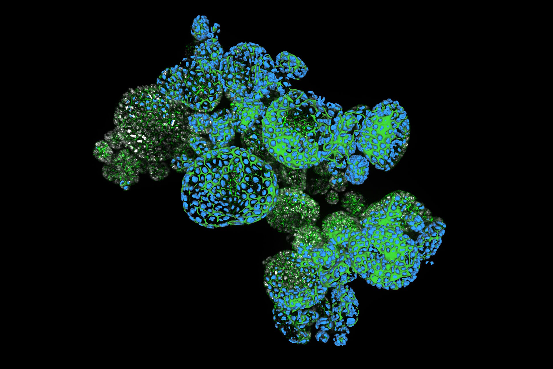
The crisp THUNDERed images, free of blur and haze, allow clear marking of signal and background. This is then the perfect starting point for AI-based object detection, segmentation, and analysis with Aivia AI image analysis software. With quantification of parameters such as growth rates, cell migration paths and velocity, and cellular interactions within the 3D environment, you can get deeper insights into your 3D cultures.
The simple workflow path from Mateo TL to THUNDER Imager and Aivia data analysis not only streamlines the process of organoid imaging and analysis but also enhances the efficiency of research endeavors. By minimizing manual intervention and leveraging THUNDER technology, you can focus on interpreting your data and formulating hypotheses, unlocking the full potential of this solution to advance the understanding of complex biological systems.

, tubulin with Cy5 (red), and nuclei with DAPI (blue). Image courtesy of Dr. Günter Giese, Max Planck Institute for Medical Research, Heidelberg, Germany.")

