
That is why Leica LMD Systems allow for collection directly into your downstream analysis containers, such as PCT microtubes for fast tissue homogenization, protein extraction, and digestion .
Pressure Cycling Technology (PCT, PressureBioSciences Inc.) uses high pressure to streamline sample preparation for mass spec analysis. PCT can process LMD samples in one hour, and larger biopsy tissue pieces in under three.
The combined techniques allow for separation of the tumor microenvironment with high precision and speed. Downstream molecular analysis of the different regions will be meaningful, since they can be analyzed separately and not as a mixture.
Download Flyer
Biomarker Discovery Workflow with Single Cell Precision
Leica's Laser Microdissection systems will improve your workflow by precisely cutting only the cells you are interested in. Everything under visual control and without contamination with surrounding tissue.
Download: Flyer Biomarker Discovery

2. Fixation and staining

Fixation and staining of tissue for LMD application can be applied according to the structures of interest. This can comprise both, brightfield and fluorescence stains.
This step can be automated with a stainer.
In general, most stains are compatible with downstream proteomics. Nevertheless, it is worth investigating before.


Mouse brain, Cresyl violet. Before and after auto detection (ADM) of neurons.
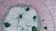
4. Laser microdissection

Next, the defined ROIs are dissected by the laser under visual control and directly collected via gravity.
Isolate cancer material or any other ROI with a few mouse clicks and pool – if necessary – in one or different reaction vessels if desired.
You can use standard consumables, such as PCR tubes. Or collect them directly into your downstream analysis vessel, e.g. PCT μTubes (Pressure BioSciences Inc.) with a dedicated LMD collection device, see next step.

5. Protein preparation

Homogenization, extraction and digestion of minute samples can be done e. g. with Pressure Cycling Technology (PCT) from Pressure BioSciences Inc. Following the LMD-PCT workflow this allows for rapid and reproducible preparation of peptides in just over one hour (Lee S et al., Cell Reports 2020), saving several hours, and providing hands off sample preparation for minute tissue samples.

6. Analysis

Analyze your samples by mass spectrometry methods and identify your biomarkers.
Typical fields of research
- Biomarker Discovery
- Cancer Research
- Personalized Medicine
- Translational Research
- Proteomics
- Metabolomics
References
- Lee S et al., Cell Reports 2020, https://doi.org/10.1016/j.celrep.2020.03.066
- Hunt AL et al., Cancer Res 2019;79 doi.org/10.1158/1538-7445.AM2019-4709
- Guo T et al., Nat Med. 2015; 21:407-13
- Shao S et al., Proteomics 2015; 15: 1-11
Related Articles
-
Selecting the Right Dissecting Microscope
You can spend many hours looking through the eyepieces of a dissecting microscope whenever…
Jan 22, 2025Read article -

Deep Visual Proteomics Provides Precise Spatial Proteomic Information
Despite the availability of imaging methods and mass spectroscopy for spatial proteomics, a key…
Dec 02, 2024Read article -

An Introduction to Laser Microdissection
The heterogeneity of histological and biological specimens often requires isolation of specific…
Oct 09, 2024Read article
Related Pages
-
Laser Capture Microdissection
Laser Microdissection, also known as LMD or LCM (Laser Capture Microdissection), is a contact- and…
Visit related page -
.")
Neuron Isolation in Spatial Context with Laser Microdissection (LMD)
After Alzheimer’s disease, Parkinson’s is the second most common progressive neurodegenerative…
Visit related page -
An Introduction to Laser Microdissection
The heterogeneity of histological and biological specimens often requires isolation of specific…
Visit related page -
 microdissected with a 10x objective (upper right). Inspection of the collection device (lower right).")
Molecular Biology Analysis facilitated with Laser Microdissection (LMD)
Extracting biomolecules, proteins, nucleic acids, lipids, and chromosomes, as well as extracting and…
Visit related page
