Contact Us

Learning the Cellular Architecture from its Optical Properties

A video presentation from Dr. Shalin Mehta at the AI Microscopy Symposium on ML and DL technologies

Analysis of anatomy and axon orientation of an adult mouse brain tissue with QLIPP. Learning_the_Cellular_Architecture_from_its_Optical_Properties_teaser.jpg

In the last 3 years, microscopists have started to use "AI based" solutions for a wide range of applications, including image acquisition optimization (smart microscopy), object classification, image classification, segmentation, restoration, super resolution and virtual staining.
The 6th edition of the AI Microscopy Symposium offered a unique forum for presenting and discussing the latest AI-based technologies and tools in the field of microscopy and biomedical imaging. Additionally, the symposium highlighted scientific breakthroughs which are enabled by machine learning or deep learning technologies.

Key Learnings

  • Quantitative label free cell imaging with phase and polarization can separate cellular structures through physical properties while providing a gentle environment for cells   
  • 2.5D UNet based deep learning models and quantitative label free imaging are computationally more efficient and almost as accurate as 3D UNets in virtual staining applications
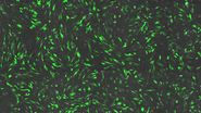
  • Cellular morphological states and transitions can be extracted from quantitative label free images using DynaMorph, a deep-learning framework

Register to view the video presentation

By clicking SUBMIT, I agree to Leica Microsystems GmbH's Terms of Use and Privacy Policy. I understand my privacy choices regarding my personal data as detailed in the Privacy Policy.

Scroll to top