Integration of Multiplexed Imaging and AI Analysis
This study explores the intricate relationships between the immune system and tumor cells in the context of cancer progression and therapy. Cell DIVE is a multiplex imaging solution capable of probing dozens of biomarkers on a single tissue section, providing a detailed molecular map of the whole tissue showcasing various cell types. Cell Signaling Technology offers IHC-validated antibodies that work seamlessly with Cell DIVE, enhancing the detection and phenotyping of various cell types in tissue samples. The integration of Aivia software simplifies spatial biology workflow by providing AI-guided segmentation, phenotyping and clustering analysis. A case study on the 4T1 murine tumor model demonstrates the utility of this spatial proteomics worfklow, revealing a heterogeneous immune landscape and complex spatial relationships within the tumor.
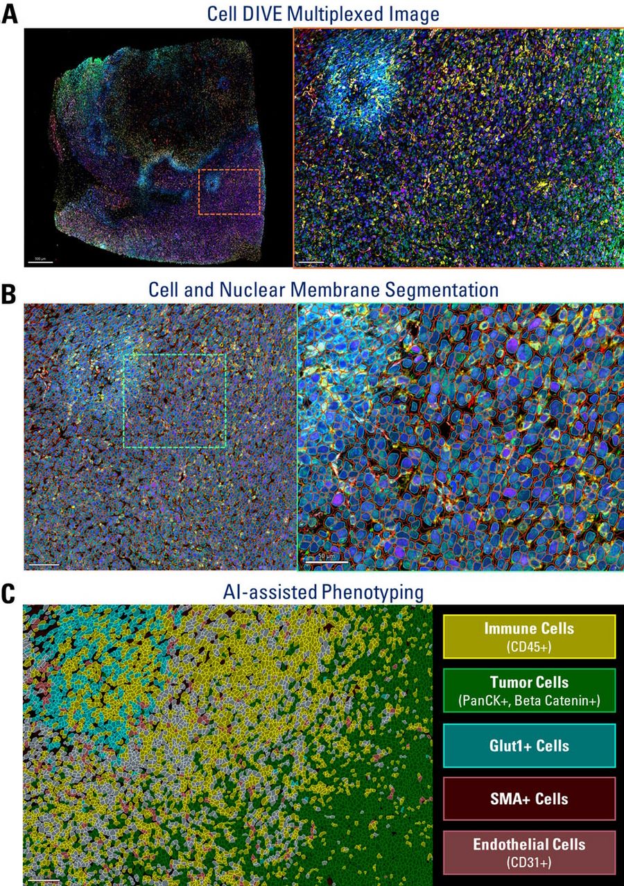
Using AI-assisted segmentaion and phenotyping on Aivia, the expression of all the markers within the Cell DIVE multiplexed whole tissue images was characterized (Figure 1).
The Impact of Tumor Microenvironment on Immune Cell Distribution
The study highlights how specific immune cell types, such as T cells, macrophages, and dendritic cells, exhibit distinct spatial distributions relative to hypoxic and necrotic regions within the tumor, suggesting the influence of the tumor microenvironment on immune cell behavior and potential implications for therapy effectiveness.
AI-powered spatial proteomics analysis using Cell DIVE and Aivia in the syngeneic 4T1 murine tumor model revealed a complex and diverse immune landscape. Overall, this study underscores the importance of combined use of multiplexed imaging and AI-powered analysis for understanding the spatial relationships and interactions within the tumor microenvironment, providing insights that could inform more effective cancer therapies.


