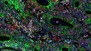
Potential of Multiplex Confocal Imaging for Cancer Research and Immunology
Expanding the frontiers of multiplex imaging - webinar on-demand

About the webinar
In this webinar, Cameron Nowell and his colleagues from Monash Institute of Pharmaceutical Sciences will share their experience in multiplex imaging and the results they were able to achieve through clever confocal imaging acquisition, as well as by utilizing other multiplexing modalities such as FLIM.
What you will learn
Key Learnings
- how multiplexed imaging can help to understand the spatial relationship between cancer cells and sympathetic nodes within the tumor and metastatic microenvironment.
- how using multiplexed imaging to visualize immune cell populations within the colon of neonatal patients with first run disease.
- how new technologies on the STELLARIS confocal platform help to extend multiplexing capabilities for life science research.
Webinar abstract
The first example comes from cancer research. It will demonstrate a unique method used in measuring spatial interactions in a tumor microenvironment of a triple-negative breast cancer.
The second example is about how to harness the power of multiplexed imaging to investigate changes to immune cell population size and location within the intestinal wall in patients who are high risk of developing a potentially lethal infection named Hirschsprung Associated Enterocolitis.
After their presentation, we will go in a detailed account of how the STELLARIS confocal platform enables researchers to extend multiplexing capabilities through new and improved technologies: the next generation of White Light Lasers and Power HyD detectors for optimized excitation and emission including the near infrared range, fully integrated lifetime-based imaging approaches (TauSense) and Stimulated Emission Depletion (STED).


