Principles of Multiphoton Microscopy for Deep Tissue Imaging

Fluorescence microscopy comes to its limits when imaging thick samples. Visible light is strongly scattered in biological tissues and fluorescence imaging is therefore restricted to an imaging depth of around 100 µm.
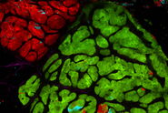
In contrast, multiphoton microscopy uses excitation wavelengths in the infrared taking advantage of the reduced scattering of longer wavelengths. This makes multiphoton imaging the perfect tool for deep tissue imaging in thick sections and living animals. Applications range from the visualization of the complex architecture of the whole brain to the study of tumor development and metastasis or the responses of the immune system in living animals.
How deep one can image with multiphoton microscopy depends on the tissue type, age of the tissue, quality of the staining but also on the excitation efficiency which is correlated to the peak power of the multiphoton laser and how well this peak power can be maintained in the imaging system.
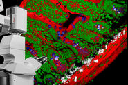
This video explains the principles of multiphoton microscopy for deep tissue imaging.


