Simplifying the Cancer Biology Image Analysis Workflow
Learn how to boost your cancer image analysis workflows with AI

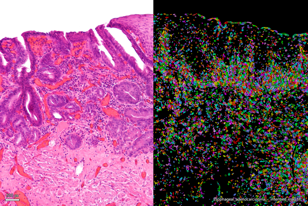
Cancer research requires large amounts of image data from 3D tissue samples or models from which information about temporal and spatial development can be extracted. As cancer biology data sets continue to grow, so do the challenges in microscopy image segmentation and quantification, making analysis highly time-consuming for researchers.
What to expect in the webinar
In this image analysis workshop, Aivia experts will cover how you can overcome these hurdles to focus on innovation and discovery in histology, and 2D and 3D cell analysis.
Key Learnings
- Conduct 2D and 3D cell analysis on a single platform
- Use machine learning to distinguish normal cells from cancerous cells
- Detect, analyze and understand the motion of cells, organelles, and particles


