Multiplexed imaging and how it compares to other imaging techniques
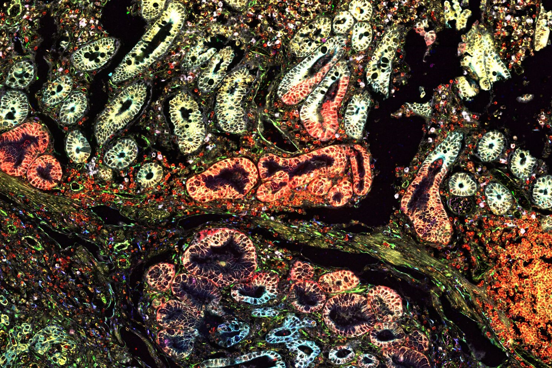
Multiplexed imaging plays a crucial role in cancer research by enabling the comprehensive characterization of tumor microenvironments. Multiplexed imaging allows researchers to simultaneously visualize various cell types and their interactions within the tumor, providing valuable insights into the tumor ecosystem. This information can help in understanding the tumor's response to therapy, predicting treatment outcomes, and identifying potential targets for immunotherapy. Additionally, multiplexed imaging facilitates the analysis of biomarker expression patterns in cancer samples. By examining multiple biomarkers simultaneously, researchers can assess the expression levels and spatial distribution of different molecules involved in cancer progression. This aids in identifying specific molecular signatures associated with different tumor subtypes, patient stratification, and predicting therapeutic response. The comprehensive analysis provided by multiplexed imaging empowers researchers to unravel the intricate complexities of cancer biology, paving the way for personalized medicine approaches and the development of targeted therapies.
In this interview, Dr. Luke Gammon, Screening Corp Facility Manager at Queen Mary University London, and Dr. David Pointu, Advanced Workflow Specialist at Leica Microsystems, discuss multiplexing imaging and how it compares to other imaging techniques. They cover the main goals of the facility, the importance of multiplexing in accessing and reusing precious samples, and the benefits it provides over traditional imaging methods. Dr. Pointu also provides insights into how multiplexing using Leica Microsystems' Cell DIVE helps produce clear tissue images at scale, using 60+ biomarkers. They also discuss their pain points, and the future potential of multiplexing.


