Key Learnings
- How to create an overview of embryos then select the ones with the right orientation and expression levels for further imaging
- How to perform a time lapse under stable conditions using the climate control functionality of Mica
- How to combine gentle widefield imaging with high resolution confocal imaging in one experiment
Speakers

Dr. Oliver Schlicker, Senior Application Manager – Leica Microsystems
Oliver received his PhD in Neuro-Cell-Biology from the University of Heidelberg, Germany. After heading the Imaging Facility at the IZI in Stuttgart, Germany he joined Leica Microsystems Wetzlar in 2008 as Application Manager, responsible for Advanced Fluorescence Widefield Microscopy.

Camilla Autorino, PhD Student – Petridou Group, EMBL Heidelberg
Camilla studies early Zebrafish embryogenesis, working on the link between tissue mechanics and specification of cell fate. She is originally from Italy, where she earned a bachelor’s degree in biology at ‘La Sapienza’ University of Rome, she then moved to Germany for her Master studies in Developmental and Stem Cells Biology at the University of Heidelberg.
The Experiment
Original broadcast date: 06th April 2022

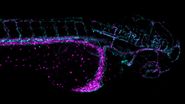
Our guest Camilla Autorino introduces us to her work, which is about studying the emergence of complexity during early phases of zebrafish development. More specifically, her focus is to analyze how changes in tissue properties like tension can impact the overall development of embryos. In this video, she demonstrates an applicational workflow, going from an overview of embryos to the selection of those with the right expression for further live cell imaging in widefield, followed by high resolution confocal imaging.
The application shows how the stable environmental control of Mica allows the further development several embryos at different stages of development in a single dish. Zebrafish embryos must be kept at 28°C; if the temperature goes higher or lower, they will develop either faster or slower, with different dynamics. This is why the absolute temperature stability and humidity control was important for this type of application.
In addition, as the embryos could be easily identified with two fluorescent markers being expressed simultaneously, the selection of the embryos with the best orientation and expression levels for later imaging steps becomes much easier. Our guest also shows how easy it is to switch from widefield to confocal with Mica, and the automated imaging setup allowed her to acquire all the necessary data sets in much fewer steps compared to conventional microscopy.


