
is mobile? false
Life Science Research
Life Science Research
This is the place to expand your knowledge, research capabilities, and practical applications of microscopy in various scientific fields. Learn how to achieve precise visualization, image interpretation, and research advancements. Find insightful information on advanced microscopy, imaging techniques, sample preparation, and image analysis. Topics covered include cell biology, neuroscience, and cancer research with a focus on cutting-edge applications and innovations.
Popular Show subnavigation
Life Science Research


Filter articles
Tags
Story Type
Products
Loading...
 where cyan indicates nuclei and magenta tight junctions.")
Rapid Check of Live Stem Cells in Cell-Culture Inserts set in Multi-Well Plates
See how efficient imaging of live iPSC stem cells within cell-culture inserts set in a multi-well plate can be done to evaluate the cells using a THUNDER Imager. Just read this article.
Loading...
 dataset, showing the biochemically distinct structures of a fresh, untreated apple slice.")
How to Prepare Samples for Stimulated Raman Scattering (SRS) imaging
Find here guidelines for how to prepare samples for stimulated Raman scattering (SRS), acquire images, analyze data, and develop suitable workflows. SRS spectroscopic imaging is also known as SRS…
Loading...

Notable AI-based Solutions for Phenotypic Drug Screening
Learn about notable optical microscope solutions for phenotypic drug screening using 3D-cell culture, both planning and execution, from this free, on-demand webinar.
Loading...

Coherent Raman Scattering Microscopy Publication List
CRS (Coherent Raman Scattering) microscopy is an umbrella term for label-free methods that image biological structures by exploiting the characteristic, intrinsic vibrational contrast of their…
Loading...
. Image courtesy of Prof. Hui Guo, School of Life Sciences, Central South University, China")
How Microscopy Helps the Study of Mechanoceptive and Synaptic Pathways
In this podcast, Dr Langenhan explains how microscopy helps his team to study mechanoceptive and synaptic pathways, their challenges, and how they overcome them.
Loading...

What is the Patch-Clamp Technique?
This article gives an introduction to the patch-clamp technique and how it is used to study the physiology of ion channels for neuroscience and other life-science fields.
Loading...

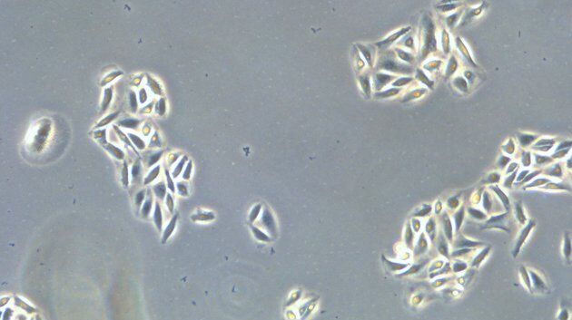
How to Determine Cell Confluency with a Digital Microscope
This article shows how to measure cell confluency in an easy and consistent way with Mateo TL, increasing confidence in downstream experiments.
Loading...

How to do a Proper Cell Culture Quick Check
In order to successfully work with mammalian cell lines, they must be grown under controlled conditions and require their own specific growth medium. In addition, to guarantee consistency their growth…
Loading...

Virtual Reality Showcase for STELLARIS Confocal Microscopy Platform
In this webinar, you will discover how to perform 10-color acquisition using a confocal microscope. The challenges of imaged-based approaches to identify skin immune cells. A new pipeline to assess…
