
Life Science Research
Life Science Research
This is the place to expand your knowledge, research capabilities, and practical applications of microscopy in various scientific fields. Learn how to achieve precise visualization, image interpretation, and research advancements. Find insightful information on advanced microscopy, imaging techniques, sample preparation, and image analysis. Topics covered include cell biology, neuroscience, and cancer research with a focus on cutting-edge applications and innovations.
Filter articles
Tags
Story Type
Products
Loading...

20 Years of Leica Laser Microdissection
Phenotype-genotype correlations are key for insight. From Eye to Insight is therefore fitting perfectly to Leica Microsystems and in particular to laser microdissection. Laser Microdissection, also…
Loading...
 - THUNDER Imager 3D Cell Culture Influenca virus – red, cilia – green, Nuclei – blue.")
How Can Immunofluorescence Aid Virology Research?
Modern virology research has become as crucial now as ever before due to the global COVID-19 pandemic. There are many powerful technologies and assays that virologists can apply to their research into…
Loading...
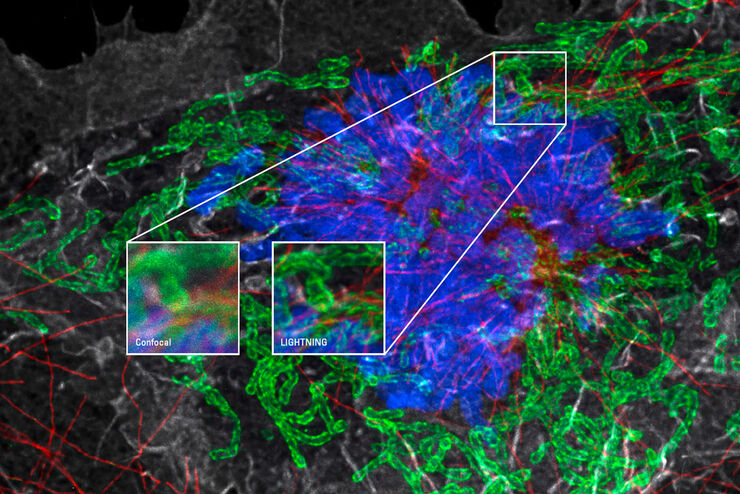
Obtain Maximum Information from your Specimen with LIGHTNING
LIGHTNING is an adaptive process for extraction of information that reveals fine structures and details, otherwise simply not visible, fully automatically. Unlike traditional technologies, that use a…
Loading...

Microscopy in Virology
The coronavirus SARS-CoV-2, causing the Covid-19 disease effects our world in all aspects. Research to find immunization and treatment methods, in other words to fight this virus, gained highest…
Loading...
Explore Innovative Techniques to Separate Fluorophores with Overlapping Spectra
In this article we explore several strategies you can take to improve the separation of fluorophores and increase the number of fluorescent probes you can distinguish in your sample.
Loading...

STELLARIS White Light Lasers
When it comes to choosing fluorescent probes for your multi-color experiments, you shouldn’t have to compromise. Now you can advance beyond conventional excitation sources that limit your fluorophore…
Loading...
TauSense Technology Imaging Tools
Leica Microsystems’ TauSense technology is a set of imaging modes based on fluorescence lifetime. Found at the core of the STELLARIS confocal platform, it will revolutionize your imaging experiments.…
Loading...

The Power HyD Detector Family
Powerful photon counting detectors on the STELLARIS confocal platform provide improved photon counting, ultra-sensitive imaging and more color options in the NIR spectrum.
Loading...
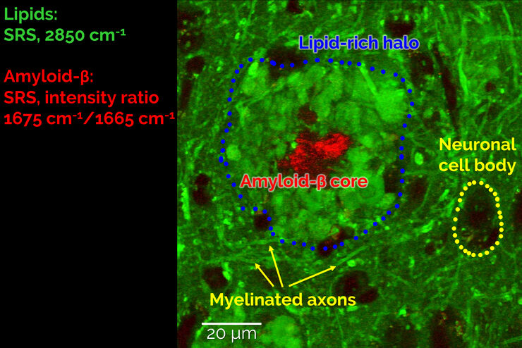
Stimulated Raman Scattering Microscopy Probes Neurodegenerative Disease
Despite decades of research, the molecular mechanisms underlying some of the most severe neurodegenerative diseases, such as Alzheimer’s or Parkinson’s, remain poorly understood. The progression of…
