
Life Science Research
Life Science Research
This is the place to expand your knowledge, research capabilities, and practical applications of microscopy in various scientific fields. Learn how to achieve precise visualization, image interpretation, and research advancements. Find insightful information on advanced microscopy, imaging techniques, sample preparation, and image analysis. Topics covered include cell biology, neuroscience, and cancer research with a focus on cutting-edge applications and innovations.
Filter articles
Tags
Story Type
Products
Loading...

Video Tutorials: Filling and Assembling of Different Carriers for High-Pressure Freezing
High pressure freezing (HPF) is a cryo-fixation method primarily for biological samples, but also for a variety of non-biological materials. It is a technique that yields optimal preservation in many…
Loading...
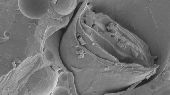
Brief Introduction to Freeze Fracture and Etching
Freeze fracture describes the technique of breaking a frozen specimen to reveal internal structures. Freeze etching is the sublimation of surface ice under vacuum to reveal details of the fractured…
Loading...
Brief Introduction to Specimen Trimming
Before ultrathin sectioning a sample with an ultramicrotome it has to be pre-prepared. For this pre-preparation, special attention must be paid to the sample size (size of the section), location of…
Loading...
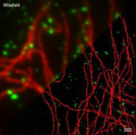
Sample Preparation for GSDIM Localization Microscopy – Protocols and Tips
The widefield super-resolution technique GSDIM (Ground State Depletion followed by individual molecule return) is a localization microscopy technique that is capable of resolving details as small as…
Loading...

Brief Introduction to Freeze Substitution
Freeze-substitution is a process of dehydration, performed at temperatures low enough to avoid the formation of ice crystals and to circumvent the damaging effects observed after ambient-temperature…
Loading...

Brief Introduction to Contrasting for EM Sample Preparation
Since the contrast in the electron microscope depends primarily on the differences in the electron density of the organic molecules in the cell, the efficiency of a stain is determined by the atomic…
Loading...

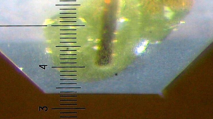
Brief Introduction to Glass Knifemaking for Electron and Light Microscope Applications
Glass knives are used in an ultramicrotome to cut ultrathin slices of samples for electron and light microscope applications. For resin and for cryo sections (Tokuyasu samples) the knife edge must be…
Loading...

Brief Introduction to Coating Technology for Electron Microscopy
Coating of samples is required in the field of electron microscopy to enable or improve the imaging of samples. Creating a conductive layer of metal on the sample inhibits charging, reduces thermal…
Loading...
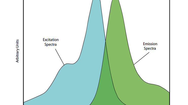
Handbook of Optical Filters for Fluorescence Microscopy
Fluorescence microscopy and other light-based applications require optical filters that have demanding spectral and physical characteristics. Often, these characteristics are application-specific and…
