How to prepare high-quality sections of polymers
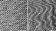
Polymers are microtomed at either room temperature or cryo temperatures, depending on their Tg. Lowering the wedge angle of the knife from 45° to 35° or even 25° results in less compression and better structure preservation. However, this also leads to a more sensitive cutting edge. An alternative approach involves oscillating the knife [1,2], which reduces compression and enhances structure preservation, particularly for rigid polymers, without introducing additional cutting artifacts.
To achieve high-quality sections, polymer samples must be properly trimmed which is best done with diamond-trimming blades. Ultramicrotomy can be performed on a dry diamond knife or with a liquid, such as a dimethyl-sulfoxide/water mixture (50/50%), keeping the temperature at -40°C so it remains below the glass transition temperature.
Successful staining of polymers
To enhance contrast when using TEM, polymers need to be stained. Common staining agents include osmium tetroxide (OsO4) or ruthenium tetroxide (RuO4). Staining can be done on the sections or the sample block after trimming. Blends may require double staining to express different phases. However, for investigating nanoparticles, nanotubes, or other inclusions, staining is unnecessary.
Obtaining smooth surfaces for AFM (atomic force microscopy) analysis
For AFM imaging, the section thickness should be kept thin (< 100nm), as sections with thicknesses in the micrometer range often do not have a smooth surface. Distinguishing between true structures and sectioning artifacts is crucial.
For ultrathin sectioning of metals, very small sample blocks (width < 50µm) are trimmed with diamond-trimming blades on trimming machines, then polished, etc. Section thickness for metals typically range from 20 to 40 nm. Some metals, like iron, cobalt, nickel, etc., can chemically react with the diamond, leading to a fast wear of the cutting edge. For such metals, it's necessary to change the diamond knife to a fresh one for each section.
For reliable AFM-image analysis, achieving a smooth section surface is crucial, as thicker sections tend to have surface patterns which are artifacts caused by the cutting process. Most brittle materials can be sectioned with 35° diamond knives, but for some hard materials, like hydroxyapatite, olivine, and certain ceramics, 45° diamond knives perform best.
Ultramicrotomy techniques for brittle materials
Ultramicrotomy is also applicable for the sectioning of brittle materials. It offers advantages, such as fast, clean, and damage-free preparation, when compared to other techniques like polishing or ion-beam milling. However, brittle materials may break into small fragments during cutting and locating the area of interest on large samples might be challenging. Target trimming machines can aid users concerning precise trimming for specific locations on the material.
Brittle-material samples are usually embedded in a rigid epoxy resin before ultramicrotomy. This method allows sections with a thickness as small as 35 nm to be obtained. For sectioning hard samples, like ceramics, semiconductors, oxides, crystals, etc., the cross section must be small (< 20µm). Alternatively, brittle-material samples can be glued onto a support with cyanacrylate glue or clamped onto an AFM sample holder. Non-embedded brittle-material samples may be sectioned as thin as 15 nm.
Conclusion
In conclusion, ultramicrotomy is a powerful preparation method for polymers, metals, and brittle materials, allowing researchers to investigate their structures and properties in high resolution with various microscopy techniques. Proper consideration of sample characteristics, knife angles, staining, and trimming techniques is essential to obtain accurate and high-quality results which can lead to useful analysis and a better understanding of these materials.
References
- C. Mayrhofer, H. Plank, H. Gnägi, I. Letofsky-Papst, Ultramicrotomy of Polymers at its best: Ultra Sonic Knives, Poster, Polymertec 2021.
- J.S.J. Vastenhout, H. Gnägi, Ultramicrotomy of Polymers Using an Oscillating Diamond Knife; Improving Polymer Morphology, Microscopy and Microanalysis (2002) vol. 8, iss. S02, pp. 324–325, DOI: 10.1017/S1431927602100560.


