Unlocking the Secrets of Organoid Models in Biomedical Research
Discover the powerful synergy of THUNDER and Spinning Disk

Wednesday 30th April 2025, 9AM EST | 2PM GMT | 3PM CET
Get ready to delve deeper into the world of organoids and 3D models, which are essential tools for advancing our understanding of human health. Navigating these complex structures and obtaining clear images for analysis can be challenging. In this event, researchers from The University of Oxford and University College London will join us to show how the THUNDER Imager Cell Spinning Disk can provide more convincing, high-quality data for deeper insights into a variety of models.
Key webinar learnings:
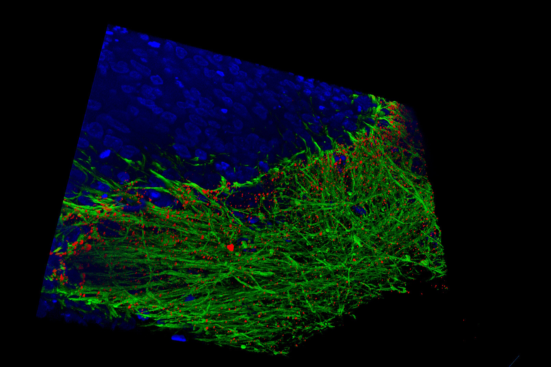
- Examining 3D human bladder microtissue infected with E.coli: Jennifer Rohn's lab at UCL has developed a pioneering 3D human urothelium organoid model (3D-UHU) to explore UTI pathology and treatment failures. Discover how clearer insights provided by the new THUNDER Imager Cell Spinning Disk can help them further explore the impact of bacterial filamentation and intracellular bacterial communities on UTI outcomes.
- Exploring human stem cell-derived mid brain organoids: The Wade-Martins group in Oxford examines disease mechanisms for Parkinson’s disease. See how they develop ventral mid-brain organoids and Enteric Nervous System (Gut-Brain axis) using stem cells and microfluidics and how much more detail they can now uncover.
- How microtubule dynamics influence cell shape: To illustrate the diversity of samples that can benefit from the synergy of THUNDER and spinning disk, we’ll also share data on individual microtubule dynamics, courtesy of the Sainsbury Laboratory in Cambridge.
This webinar is a must-attend for researchers looking to gain deeper insights into their organoid and 3D models or enhance their live cell imaging capabilities.
About THUNDER Imager Cell Spinning Disk
While spinning disk alone provides fast, versatile, confocal imaging and powerful optical sectioning, the synergistic pairing with THUNDER eliminates residual out-of-focus light, resulting in greater contrast and resolution.
This enables you to extract more statistically significant data from diverse 3D samples in less time and with reduced phototoxicity.
Typical applications include live cells, 3D cell cultures, organoids, spheroids, model organisms, plant biology and thick tissue samples.
Related Articles
-
 applied. Image courtesy of Samuel East, Uncommon Bio.")
Designing the Future with Novel and Scalable Stem Cell Culture
Visionary biotech start-up Uncommon Bio is tackling one of the world’s biggest health challenges:…
Mar 11, 2025Read article -

Mica: A Game-changer for Collaborative Research at Imperial College London
This interview highlights the transformative impact of Mica at Imperial College London. Scientists…
Feb 10, 2025Read article -

A Guide to Model Organisms in Research
A model organism is a species used by researchers to study specific biological processes. They have…
Feb 04, 2025Read article
