A Practical Guide to Virology Research

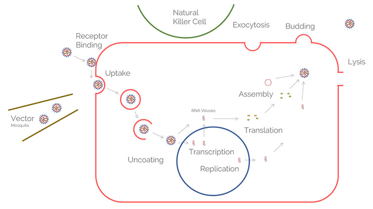
Leica solutions for imaging and sample preparation help you with the investigation of viral entry and fusion, genome integration, viral replication, assembly, and virus budding.

 Influenza_lung_porcine_ICC_3-2.jpg

The study of virus-infected tissues and cells to understand infection mechanisms and develop treatments for disease has dramatic implications for human health. Leica solutions for imaging and sample preparation help you with the investigation of viral entry and fusion, genome integration, viral replication, assembly, and virus budding. Data from analysis of well-prepared specimens also are important for studying the related cellular mechanisms and immune responses. The ultimate goal is to develop intervention strategies.

Viruses are investigated from the biopsy to single virion level. Some of the most common viruses studied are influenza, coronaviruses (SARS-CoV-2 causing COVID-19, etc.), herpes, hepatitis, dengue, zika, HIV, rabies, and Ebola, but there are many others.

Host Side of the Virus Infection Process

For virology research model organisms are sometimes used, however, they do not mimic well the human host morphology. Hence, researchers more often rely on human tissue, biopsies, or cell culture studies.

Ex vivo infection models can be generated from tissue, biopsies, or animal models. These models can be investigated with the aid of Leica stereo microscopes.

Cell cultures, spheroids, and organoids can be monitored with the help of Leica widefield (compound) microscopes.

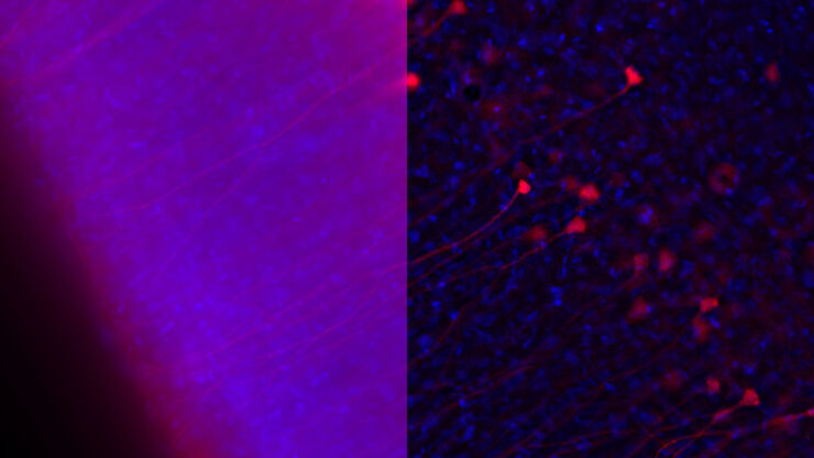
Further initial investigations of infection models, 2D or 3D cell cultures, are achieved with more sophisticated Leica fluorescence microscopes, such as THUNDER imagers.

More reliable downstream genetic analysis can be done with laser microdissection from Leica Microsystems which isolates specific cells from surrounding tissue.

Related articles

Microscopy in Virology

The coronavirus SARS-CoV-2, causing the Covid-19 disease effects our world in all aspects. Research to find immunization and treatment methods, in other words to fight this virus, gained highest…
Image source: Adobe Stock

How to Sanitize a Microscope

Due to the current coronavirus pandemic, there are a lot of questions regarding decontamination methods of microscopes for safe usage. This informative article summarizes general decontamination…

High Resolution Fluorescence Imaging for Virology Research

High resolution microscopy is an asset for infectious disease studies as it allows you to track causative viruses or pathogens. Combined with fluorescent labelling of cellular or viral components, it enables you to probe multiple aspects of infection processes.

Thus, both laser scanning confocal and total internal reflection fluorescence (TIRF) microscopy obtaining super-resolution are instruments of choice for viral research.

Advanced microscopy techniques, such as multiphoton and light sheet microscopy, can give you access to infection events occurring under different contexts, such as tissue sections, animal models, and 3D infection models like organoids.

Leica microscopes offering you advantages for the study of 3D specimens concerning virology research include the STELLARIS confocal platform and TIRF solutions.

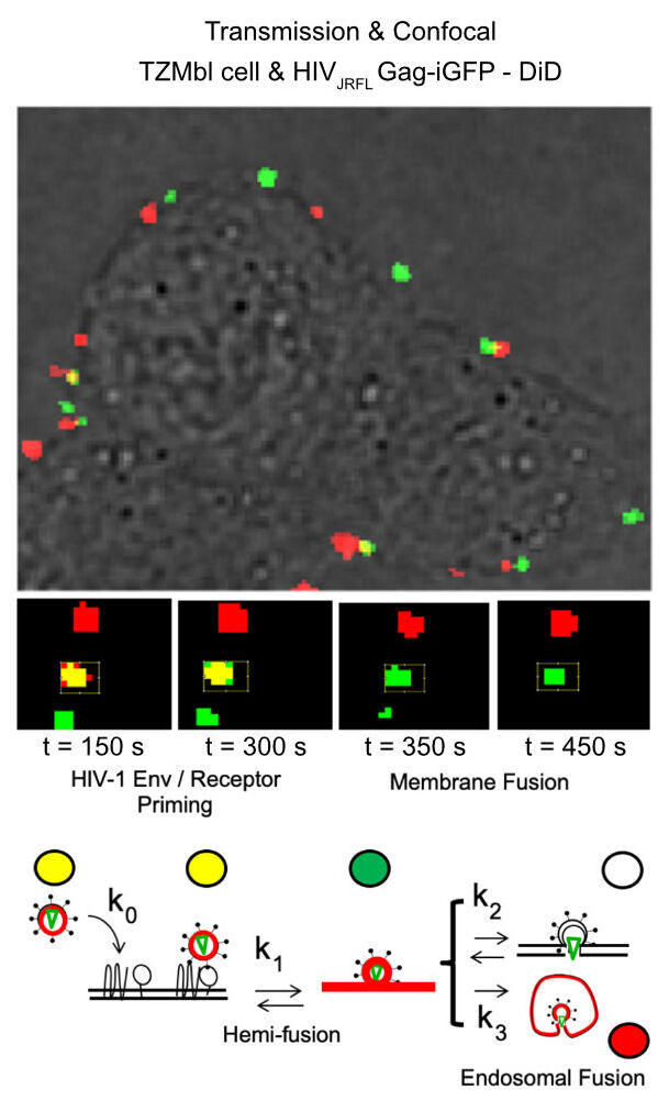
Image and diagram adapted from Coomer et al. PLOS Pathogen (2020) 16(2): e1008359, DOI: 10.1371/journal.ppat.1008359

Related articles

Molecular structure of the green fluorescent protein (GFP)

Introduction to Fluorescent Proteins

Overview of fluorescent proteins (FPs) from, red (RFP) to green (GFP) and blue (BFP), with a table showing their relevant spectral characteristics.
Cellular microtubule network in a fibroblast cell.

How to Prepare your Specimen for Immunofluorescence Microscopy

Immunofluorescence (IF) is a powerful method for visualizing intracellular processes, conditions and structures. IF preparations can be analyzed by various microscopy techniques (e.g. CLSM,…
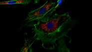
Image of immunofluorescently labelled cells where mitochondria are indicated with red, nuclei with blue, and actin with green.

Studying Virus Replication with Fluorescence Microscopy

The results from research on SARS-CoV-2 virus replication kinetics, adaption capabilities, and cytopathology in Vero E6 cells, done with the help of fluorescence microscopy, are described in this…
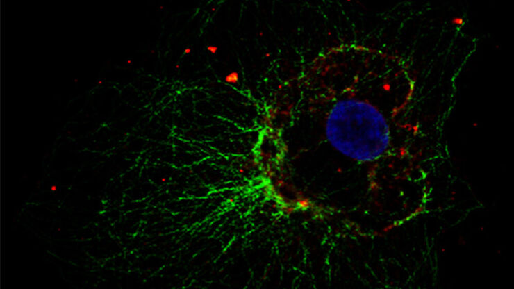
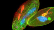
Influenca in lung epithelial cells (porcine) - THUNDER Imager 3D Cell Culture Influenca virus – red, cilia – green, Nuclei – blue.

How Can Immunofluorescence Aid Virology Research?

Modern virology research has become as crucial now as ever before due to the global COVID-19 pandemic. There are many powerful technologies and assays that virologists can apply to their research into…

Ultrastructure of Viruses

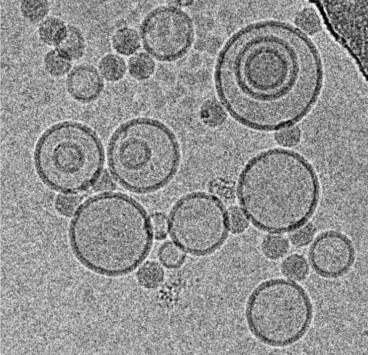
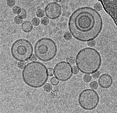
For the evaluation of virus ultrastructure, you need a resolution approaching 1 nm. At present, this resolution can only be achieved with electron microscopy. Cryo EM can achieve even sub-nanometer resolution.

Ultrastructural resolution allows you to actually see the host and virus interaction at the nanoscale, as well as confirm the results from different analytical technologies and identify potential drug target sites. Furthermore, ligands or small molecules of cell surface receptors targeted during viral particle interactions are key to the structure-guided design of vaccines and viral entry inhibitors.

For conventional and cryo EM, the sample preparation is crucial. It requires special equipment and expertise which Leica Microsystems offers you via an extensive portfolio for EM sample preparation as well as the advice from Leica experts.
Related articles

Exploring the Structure and Life Cycle of Viruses

The SARS-CoV-2 outbreak started in late December 2019 and has since reached a global pandemic, leading to a worldwide battle against COVID-19. The ever-evolving electron microscopy methods offer a…

Immersion Freezing for Cryo-Transmission Electron Microscopy: Applications

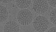
A well established usage case for cryo-TEM is three-dimensional reconstruction of isolated macromolecules, virus particles, or filaments. On one hand, these approaches are based on averaging of…
Scroll to top