Discover the anatomy
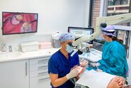
In the below video, Dr. Gorni demonstrates how he performs an inspection of the root canal of a lower molar with his dental microscope to accurately understand anatomical details of the root canal system. The use of the dental microscope provides enhanced visualization benefits, enabling a precise investigation of the anatomy of the molar and revealing additional canals.
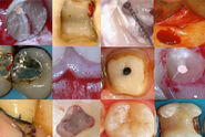
The mesial root of this lower molar anatomy is quite complex. In most cases, there is an additional canal starting from the isthmus. In this specific case, it is located directly between the mesiobuccal and the mesio-lingual canal. The canal system is very small and is hidden under a small portion of hard tissue. Using a small ultrasonic tip, Dr. Gorni reveals the orifice. The microscopic view allows a very precise cutting, helping to preserve the anatomy.
Cleaning and shaping the root canal system
The most important part of root canal treatment is the careful cleaning. Performing ultrasonic irrigation is therefore an important step. With a specific ultrasonic tip, the irrigation liquid is agitated to reach deep areas inside the root canal system for efficient cleaning. This video, taken at high magnification with the M520 F20 dental microscope, shows how deeply the activated fluid penetrates and cleans the canal.
Invasive cervical resorption (ICR)
In this case of a second upper molar with an invasive cervical resorption, see how important it is to work under high magnification with a dental microscope to ensure that all pathological tissue is properly removed in the affected area. After cleaning the area, the dental surgeon was able to determine whether the tooth was recoverable. In this case, the defect of the tooth could be repaired with bioceramic cement.
Lower molar retreatment
This case showcases removal of existing restorations in a lower molar that needed to be treated again because previous treatments were inadequate, with parts of the pulp tissue still present. The disassembling in this lower molar retreatment is quite simple. There is copious bleeding, and the root canal system is not shaped properly. Some of the pulp tissue is in the isthmus of the mesial root. After the shaping of the endodontic space, the root canal is completely empty and clean. These steps performed by Dr. Gorni led to successful treatment.
Broken file removal
In this video, you can see how Dr. Gorni precisely removes broken file fragments with an ultrasonic tip. This is a typical example where a fragment of a rotary file is found in the mesial root of a lower molar. Broken files that are located under the orifice, especially in the middle or lower third of the canal, cannot be removed without a microscope. Good visualization is required to address this challenge. With the microscope, the dentist can be very precise while cutting the tissue around the broken file fragments, using an ultrasonic tip. The ultrasonic energy removes tooth structure and vibrates the fragment loose.
Disassembling in a retreatment case
This video demonstrates dental microscope benefits for retreatment and the removal of gutta-percha, which are some of the most challenging tasks in endodontics surgery. When working in small canal spaces, there is a potential risk of damaging the dentine walls. The improved visibility with the dental microscope allows a thorough control of the use of the instruments. At the end of the disassembling, the root canal system is completely empty and ready to be filled again.
Microsurgery: Endodontic flap
A precise surgical incision and flap design are important to any endodontic surgery. Under high magnification, the incisions can be made very accurately. According to Dr. Gorni, the precise flap design also supports the healing of the soft tissue. In this short video, you will see how Dr. Gorni uses microscopic visualization to achieve this goal.
Sinus lift
The use of a dental microscope is beneficial for many dentistry procedures, but especially for complex endodontic surgeries. This includes sinus lift, a complex and difficult procedure which requires high precision, especially when the Sneider membrane must be risen. This thin and delicate membrane covers the entire cavity of the maxillary sinus. It is vital to obtain a detailed view using a dental microscope, as it supports the surgeon in gently managing the sensitive membrane and avoiding damages. A perforation of the membrane would be dangerous and could lead to treatment failure. The use of a dental microscope can help to reduce damage and complications during and after the procedure.
In this video, you can see how Dr. Gorni benefits from improved visualization when working on the delicate and thin Sneider membrane, and how it stayed intact, and the bone replacement filling was put into place.
Discovering the anatomy during micro endodontic surgery
During endodontic procedures, such as retrograde root canal treatment, the dental microscope provides the necessary light and magnification to enable the endodontist to better see and treat the different areas. Accuracy is key in retrograde endodontic treatment, and the microscope supports the surgeon in careful retro-prep and precise root end filling. The entire root canal anatomy can be checked reliably, with the microscope view revealing lateral canals and extra exit portals. This helps avoid a leakage in the final obturation of the space.
The benefits of a dental microscope in micro endodontic surgery
Dental microscopes have numerous advantages for micro endodontic surgery according to Dr. Gorni. First, they enable minimally invasive procedures and as such play an increasingly important role. Endodontists can work with high precision from the first cut to the suture, thanks to a detailed and bright microscope view. The osteotomy and target search also become easier, as endodontists can easily control the microscope’s visualization. Apical resection, root end preparation and root end filling are also simpler to perform. For the differentiation of canal anatomy based on endodontic principles, it is essential to have a clear, deep view allowing to see everything down to the smallest detail.
In all these endodontic and micro-dentistry procedures, the microscope provides a detailed view, allowing the surgeon thorough control of the instruments and ensuring all pathological tissue is properly removed to preserve the tooth. According to Dr. Gorni, a microscope supports him to carry out individual treatment steps more precisely and carefully which may improve the success of the treatment. He therefore believes that the dental microscope has become indispensable for successful endodontic surgery.
Disclaimer: Please note that off-label uses of products may be discussed. Consult with regulatory affairs for cleared indications for use in your region. The statements of the healthcare professionals included in this presentation reflect only their opinion and personal experience. They do not necessarily reflect the opinion of any institution with whom they are affiliated or Leica Microsystems.
Suggested reading
- S. A. Mallikarjun, P. R. Devi, A. R Naik, S. Tiwari, Magnification in dental practice: How useful is it? JHRR (2015) vol. 2, iss. 2, page 39-44, DOI: 10.4103/2394-2010.160903
- R. Hegde, V. Hegde: Magnification-enhanced contemporary dentistry: Getting started. JID (2016) vol.6, iss.2, page 91-100, DOI: 10.4103/2229-5194.197695
- U. K. Das, .S. Das: Dental Operating Microscope in Endodontics-A Review. IOSR Journal of Dental and Medical Sciences (2013) vol. 5, iss. 6
- G. B. Carr , C. A. F. Murgel: The use of the operating microscope in endodontics. Dent Clin North Am (2010 Apr);54(2):191-214.doi: 10.1016/j.cden.2010.01


