Key Learnings
Prof. Schmidt explains that GLOW800 offers more information compared to conventional ICG angiography as well as increased speed, safety and convenience. With GLOW800, the surgeon is much more confident. It also allows to use a lower ICG dose, which is beneficial for the patient, and enables repetition if needed.
In the below videos, Prof. Schmidt discusses these benefits. In the first video, he addresses efficiency and ICG dose management with GLOW800. In the second one, he shares the advantages during procedures such as aneurysm and arteriovenous malformation (AVM) surgeries. In the third video, Prof. Schmidt highlights how GLOW800 supports teaching and OR staff communication. Discover the three videos and their full transcripts.
For more information about GLOW800 Augmented Reality fluorescence or how to choose a neurosurgery microscope, contact a Leica representative. Our team will gladly answer your questions and help you find the right solution for your needs.
Transcription Video 1
“GLOW800 is helping us in a way that it incorporates morphological or anatomical information, which you can directly visualize on the microscope. So, the benefits of using GLOW800 is that it’s faster, you get the information faster than previously because you have it directly in your visual field, you don’t have to look around, watch the video over and over.
It makes the procedure safer, because you are able to manipulate during the ICG angiography, while using GLOW, you can manipulate in your surgical view and that helps a lot to get more information. And it improves the confidence while you’re doing the surgery.
It’s certainly faster than the conventional way to do it. It’s simpler to use than the conventional way to do the ICG angiography. And that’s very convenient, it gives you more information, it gives you more confidence during surgery.
With certain systems, we need a little bit more of the ICG. Using the Leica microscope and the GLOW800 system, we use a very low dose and we can repeat it without problems, and that works very well.
GLOW800 is actually very sensitive. In complicated cases, it gives you an advantage that you just use less, or you repeat it more often, and you have a good feeling about doing it.”
Transcription Video 2
“Today we performed microsurgery on a 46-year old female. She has an aneurysm. It was an incidental finding; it was a very broad based one and with a size of 9mm at the Acorn position. Therefore, the indication was to use a microsurgical clipping.
It went very well, we used a frontobasal approach and after preparation of the A1 segment from the right side, we were able to clip it without any temporal clipping and we confirmed the exclusion of the aneurysm by using GLOW800.
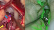
We regularly use ICG fluorescence angiography. Now, with the introduction of GLOW 800, it can be incorporated to the neurosurgical procedure much more fluently, because you can view it inside the microscope. It has false colors, and we see the anatomical relation of the aneurysm and of the fluorescence, which we were not able to see while we were using the conventional ICG. So, it’s much more integrated into the normal flow of microsurgery.
We use ICG fluorescence for all our neurovascular cases, for the blood flow and also some information on whether the aneurysm if fully excluded from the circulation. And GLOW800 is helping us to do this.
We also use GLOW800 for other vascular cases than aneurysm surgeries or microsurgical excision of arteriovenous malformations. We use it if we do bypass surgery and I’ve even used it during surgical removal of some vascular tumors, like hemangioblastoma.
It’s simpler to use than the conventional way to do the ICG angiography. And that’s very convenient, it gives you more information, it gives you more confidence during surgery.”
Transcription Video 3
“There is one aspect which is very important with GLOW800 or the way it visualizes. If you show it to students, if you show it to residents, for teaching purposes, it’s perfect. Because you have all the information together, you can explain it. You have the information and can repeat it and demonstrate all the different aspects during the surgical procedure.
And that’s very useful, because with the conventional black and white images of the ICG angiography, it’s very hard for beginners or students to follow all the aspects of the microsurgical procedure, and all the information you get with it.
The team appreciates it very much, because everyone can understand it if it’s on the screen, and you see all the colors with the anatomical information. That is something that leads to everyone immediately picking up what’s going on there.”
About GLOW800 Augmented Reality fluorescence
GLOW800 Augmented Reality fluorescence and ICG allow to observe cerebral anatomy in natural color, augmented by real-time vascular flow, with full depth perception.
It provides one augmented view during vascular neurosurgery for enhanced confidence during aneurysm clipping, AVM removal, microvascular decompression or bypass surgery for example.
GLOW800 Augmented Reality fluorescence can be viewed directly in the eyepieces of the Leica Microsystems M530 OHX neurosurgery microscope. It is also available on the Leica ARveo digital Augmented Reality microscope.
For more information about GLOW800 Augmented Reality fluorescence or Leica neurosurgery microscopes, contact a Leica representative.
The statements of the healthcare professional included in this article reflect only his opinion and personal experience. They do not necessarily reflect the opinion of any institution with whom he is affiliated.


