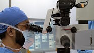
First clinical case: Standard cataract surgery & standard phacoemulsification
In this standard cataract case, the surgical plan was to perform standard phacoemulsification with a 2.2mm incision. For this type of case, Dr. Moraru uses either the binocular M844 microscope viewing system or a 3D visualization system.
As such, the surgeon has three visualization possibilities: the intraoperative OCT screen which is in front of the surgeon, a picture-in-picture view on the 3D visualization screen or a dual image through a split screen or two different screens.
This offers numerous advantages for cataract surgery teaching, as well as for scientific purposes helping to observe wounds architecture, phaco tip position, fluctuations of the anterior chamber depth, zonules behavior, etc.
Enhanced visualization is also valuable in unconventional or complex cataract surgery cases such as mature cataract with no red reflex, posterior polar cataract, posterior capsule rupture (with or without drop nucleus/cortex), zonular rupture, subluxated lens, small pupil and Intraoperative Floppy Iris Syndrome (IFIS).
In a mature cataract case with a relatively small pupil, the intraoperative Optical Coherence Tomography picture-in-picture image helped visualize the position of the posterior capsule.
Second Clinical Case: Advanced Cataract Surgery & Complex Phacoemulsification
In this case, a 62-year-old male patient presented with advanced cataract in the right eye, with finger-counting visual acuity. He didn’t have any other abnormal eye pathology. The surgical plan was to use standard chop phacoemulsification before posterior chamber intraocular lens (PC-IOL) implantation.
However, there were complications during surgery. At the very end of the procedure, when attempting to hydrate the paracentesis, the surgeon noticed a strange thin line that was slightly curved and thought it could be an accidental Descemet membrane detachment.
Learn more and discover three other cases including complex cataract surgery cases. Enter your information to download the full case study.
Please note that off-label uses of products may be discussed. Please check with regulatory affairs for cleared indications for use in your region. The statements of the healthcare professionals included in this clinical case reflect only their opinion and personal experience and not those of Leica Microsystems. They also do not necessarily reflect the opinion of any institution with whom they are affiliated.


