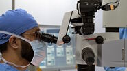
Proveo 8 is an ophthalmic microscope supporting a smooth and uninterrupted surgical workflow during anterior and posterior segment surgery. The FusionOptics technology allows to see more detail in one view. There is no need to constantly refocus. The microscope includes a stable red reflex for cataract surgery, offering consistent images throughout.
Key Learnings
Prof. Spitzer explains the advantages of the Proveo 8 ophthalmic microscope and how it helps achieve better outcomes and increase safety.
- In the first video, he shares the features that make Proveo 8 an ideal solution: excellent imaging quality, an outstanding depth of field, the red reflex and easy handling.
- In the second video, he explains how Proveo 8 supports the surgical workflow, in a very intuitive manner.
- In the third video, he describes the benefits of Proveo 8 when it comes to teaching, training and teamwork.
The statements of the healthcare professional included in this article reflect only his opinion and personal experience. They do not necessarily reflect the opinion of any institution with whom he is affiliated.
Transcription of Video 1
Overcome Surgical Challenges in Ophthalmology with the Proveo 8 microscope
“It is a challenge that we often have surgical procedures that involve both the anterior and the posterior segments, meaning combined surgeries, for example severe injuries that affect both the anterior and posterior segments of the eye, or just combined vitrectomies that require lens extraction at the same time.
In such situations, you want a good microscope, such as the PROVEO 8 from Leica, which is suitable for both types of procedures, because it features very good imaging and handling properties, both for the front and rear section.
The core qualities of the product, such as fine imaging quality, good red reflex, easy handling and easy cleaning options of the microscope are very good. Now, after more than half a year, we are very happy to have our Leica PROVEO 8 microscopes.
What we found particularly appealing was the very high resolution, the good depth of field of the microscope, and what I find truly impressive in the product is its very good camera – a wonderful HD quality camera.
With this camera, you can make very nice video recordings including outstanding display on the monitor; both on the monitors that came with the microscopes and on the monitors that we have in our operating room anyway.
I find that the depth of field of the PROVEO 8 is outstanding. The red reflex is really fantastic and proves to be a particular advantage, especially in cataract surgery. This was a huge step forward compared to our earlier microscopes. This made us realize that within the last few years, something has changed enormously for the better.
To begin with, the red reflex is very pleasant and also very stable even if you are somewhat off-center with the microscope, with the X/Y control, the red reflex is still extremely good and doesn’t get lost that fast. This is what I appreciate in cataract surgery.
I find the IOL compass very convenient when implanting toric lenses because it allows me to check precisely how the axis is positioned. I feel it is superior to a mark with the pendulum corneal marker because here, I obtain a continuous line, whereas the markings often dissipate with the corneal marker and I am never quite sure how well they are placed. I feel this is very convenient, and I also welcome the fade-in and fade-out of the templates.
About the motorized BIOM, I especially like the fact that it automatically generates a re-inverted image and is capable of calling up one’s own presets automatically. So you don’t have to refocus that much. The imaging properties are good, and we have various magnifying lenses at our disposal. The widefield lens takes me far in the periphery; with the macula lens, peeling becomes easy. What is nice compared to a Kilp lens is that I have quite a decent view of the periphery when peeling.
Another pleasant feature in the motorized BIOM is its synchronized focus, in other words, the distance between the front lens and the cornea always remains the same while I am focusing. This reduces the risk of touching the cornea.
The better image quality makes it naturally more comfortable for the surgeon to operate. It enhances the safety of the patient. The better the imaging quality and the image stability of the microscope, the safer the operation becomes and the better its outcome will be.”
Transcription of Video 2
Ophthalmology: Facilitate your Surgical Visualization, Workflow & Service with the Proveo 8 microscope
“My name is Martin Spitzer and my focus area is retinal diseases. But generally, I perform almost all forms of intraocular procedures. Every year, that makes about 400 vitrectomies, 500 cataract procedures, a few dozen glaucoma procedures – which are not my primary specialty – as well as a few cornea transplantations and minor procedures. My focus area is retinal surgery and retinal diseases.
It is a challenge that we often have surgical procedures that involve both the anterior and the posterior segments, that is combined surgeries, for example severe injuries that affect both the anterior and posterior segment of the eye, or just combined vitrectomies that require lens extraction at the same time.
In such situations, you want a good microscope, such as the PROVEO 8 from Leica, which is suited to both types of procedures, because it features very good imaging and handling properties, both for the anterior and posterior segment.
Since very recently, we have been using the PROVEO 8 from Leica. We are very satisfied with it. In total, we have acquired three of these Leica PROVEO 8 microscopes. One of them is equipped with an intraoperative OCT (EnFocus) from Leica. Furthermore, we have one system with an IOL compass, which we use for toric lenses, special lenses or when we implement, say, toric multifocal lenses in a femto cataract surgery.
The particularly outstanding feature of Leica’s PROVEO 8 is its intuitive usability. It is almost self-explanatory how to use the handles and footswitches. The microscope is very easy to use and after a brief instruction, you feel very comfortable with it. This applies not only to the surgeon but also to the surgical team.
We found the transition from our earlier microscope to the PROVEO 8 convenient and easier than expected, as I must admit, we looked at a number of new microscopes to find a solution that combined very good imaging quality, good user-friendliness and a reasonable price-performance ratio. This ultimately led to Leica getting the contract. So to speak, Leica beat the others in this race. Over the years, I have also experienced the support of other manufacturers and I must say, I am really impressed with Leica. In my opinion, they are among the best.”
Transcription of Video 3
Ophthalmic Surgery: Enhance Teaching & Teamwork with the Proveo 8
“My name is Martin Spitzer and my focus area is retinal diseases. But generally, I perform almost all forms of intraocular procedures. Every year, that makes about 400 vitrectomies, 500 cataract procedures, a few dozen glaucoma procedures – which are not my primary specialty – as well as a few cornea transplantations and minor procedures. My focus area is retinal surgery and retinal diseases.
The microscopes from Leica are all well suited for training. For all microscopes, we have acquired fully equipped PROVEO 8 all with assistant binocular tubes, which gives the assistants the same view as the main surgeon, with side assistant microscopes for a same view as the main surgeon.
In addition, we have very nice monitors in our ORs. All microscopes are equipped with large HD monitors in all three operating rooms. We enjoy the image quality of the camera. For our surgical team in particular, this was a great experience because they were able to see on the monitor for the first time, how beautiful ophthalmic surgery is.
Previously, the image was always quite blurry, because we didn’t have such a good camera. Additionally, we have connected one of the PROVEO 8 to the 3D system. So, the surgical team can see the surgery with 3D glasses just as clearly as the main surgeon, who is operating heads-up with 3D glasses as well. Furthermore, with 3D glasses and a heads-up display, this allows the surgeon to perform the operation without using the eyepieces.
It’s only a small portion of 3D heads-up surgeries that we do, but as a university clinic, we have students and assistant doctors in training and young specialists being trained as intraocular surgeons and for such purposes, this system is excellent.
It is a particular advantage that we work with our current microscopes in parallel. On the one hand, we can perform surgery thanks to the 3D heads-up display; on the other hand, at the same time, it offers the opportunity of looking through the eyepieces right after. You don’t have to change the set-up in between.
We integrate this system to the education of young doctors who are trained as ophthalmologists, but also in particular into the education of our medical students. They thus get a completely different impression of ophthalmic surgery than if they view it in two dimensions on the monitor only.
I believe that the Leica PROVEO 8 is very well suited as an educational microscope.”


