Pioneering in corneal surgery with intraoperative OCT
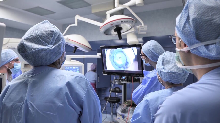
Prof. Luigi Fontana, Director of the Ophthalmology Unit at St. Orsola Hospital, University of Bologna, Italy, has been an early adopter of the EnFocus intraoperative Optical Coherence Tomography (OCT) from Leica Microsystems. This technology allows surgeons to see what lies underneath the surface and how tissue reacts to surgical maneuvers during eye surgery.
Prof. Fontana now routinely uses EnFocus intraoperative OCT in almost all his corneal transplant procedures, given the benefits he has experienced, which he outlines below.
1. Ergonomics and compact design: Enhancing surgeon comfort
One of the standout features of the new Proveo 8 with integrated intraoperative OCT is its compact scan head design. According to Prof. Fontana, “The surgeon can fit their legs under the patient bed more easily and doesn't have to strain their back to reach the eyepieces.” This ergonomic improvement represents a significant advancement from Prof. Fontana’s perspective.
2. High-resolution images: See the fine details and layers
With this technology, corneal surgeons benefit from high-resolution intraoperative OCT-imaging. Prof. Fontana points out: “Resolution in surgery is everything. We need to be able to see fine details of the structures that we operate on. Resolution must allow the surgeon to see fine details, like all the cornea layers that we excise and isolate."
This is particularly important and common across corneal transplant methods. Prof. Fontana shares a few examples: “For instance, in Descemet membrane endothelial keratoplasty (DMEK), being able to see how the donor roll is actually shaped, and its orientation, is fundamental in order to conduct the surgery in the best possible way. In deep anterior lamellar keratoplasty (DALK) with the big bubble technique, resolution allows the surgeon to determine the position of the cannula inside the cornea in order to inject air in the right position.” As such, “resolution has a major role in determining the application of intraoperative OCT to lamellar corneal transplantation.”
3. Extended scan depth: View of the entire anterior chamber
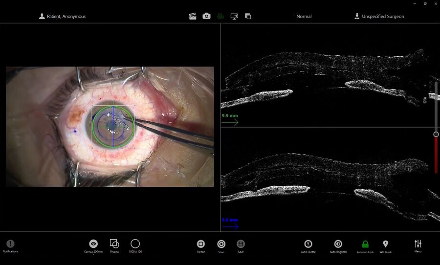
The EnFocus intraoperative OCT built in the Proveo 8 microscope offers enhanced visualization during corneal surgery. Prof. Fontana explains: “In one frame, you can see the cornea in its entire thickness as well as the anterior chamber, the angle of the iridocorneal angle as well as the iris plane and also the surface of the lens”.
One of the major benefits of EnFocus intraoperative OCT, according to Prof. Fontana, is the scan depth which gives the ability to “make accurate measurements of the layer that we are treating”, in particular, the thickness of the cornea. The real-time assessment provides enhanced insights and supports decision-making. Prof. Fontana says: “We are able to picture the entire cornea using radial scans and analyze the thickness of the cornea at all degrees”. He believes this supports the repeatability and safety of the procedure, allowing to avoid some complications that may occur during surgery.
4. Advantages for teaching
The EnFocus Intraoperative OCT is also an educational and teaching tool. As an example, Prof. Fontana organized a learning session for corneal surgeons to share the latest developments in anterior lamellar keratoplasty and posterior lamellar keratoplasty.
“The observer surgeons were remotely connected to the main operating room, observing surgery from another room. And we were able to exchange opinions during the surgery. It is quite a revolution that observer surgeons can now also view sectional images in a split-screen view on the monitor in addition to the white light microscope view.” He also highlights: “This was very useful from a technical point of view, as we were able to demonstrate to them the advantage of introducing a new technology to the standard microscope.”
Why Prof. Fontana chooses EnFocus intraoperative OCT system from Leica?
Prof. Fontana has evaluated several intraoperative OCT systems available on the market. He stated,
I found the EnFocus intraoperative OCT from Leica Microscope to meet our needs very well in terms of image quality, scan depth, and ergonomics. The system aligns well with our requirements and the needs of our team.
Conclusion: Intraoperative OCT is a necessity for corneal surgery
Intraoperative OCT enhances insights during cornea transplant procedures. In real-time surgeons get additional information via a high-resolution OCT image, showing how the subsurface tissue reacts to their surgical maneuvers. Intraoperative OCT imaging eases the assessment of the fine layers of the cornea, which is key for all methods of corneal transplants. It can help surgeons to overcome uncertainties and, avoid potential complications. According to Prof. Fontana: “We can assure you that the pre-operative measurements we took are confirmed during the operation. This is highly relevant to avoid potential complications that may arise during deep anterior lamellar keratoplasty.”
Prof. Fontana says: “I recommend intraoperative OCT because it's basically the applications of OCT that we use routinely in the clinic for the surgical treatment of both anterior segment and posterior segment disease.” In addition, the EnFocus intraoperative OCT has unique features supporting smooth and efficient surgery. He particularly enjoys “the quality of the image acquisition, the scan depth, the user friendliness, and the ergonomics.”
Watch the full video from Prof. Fontana to learn more.
Disclaimer
The statements of the healthcare professionals included in this presentation reflect only their opinion and personal experience. Not all products or services are approved or offered in every market and approved labeling and instructions may vary between countries. Please contact your local Leica representative for details.


