
Medical Specialties
Medical Specialties
Explore a comprehensive collection of scientific and clinical resources tailored for HCPs, including peer insights, clinical case studies, and symposia. Designed for neurosurgeons, ophthalmologists, and specialists in Plastic and Reconstructive surgery, ENT, and dentistry. This collection highlights the latest advancements in surgical microscopy. Discover how cutting-edge surgical technologies, such as AR fluorescence, 3D visualization, and intraoperative OCT imaging, empower confident decision-making and precision in complex surgeries.
Filter articles
Tags
Products
Loading...

Tissue Image Gallery
Visual analysis of animal and human tissues is critical to understand complex diseases such as cancer or neurodegeneration. From basic immunohistochemistry to intravital imaging, confocal microscopy…
Loading...
 and astrocytes (green) in a cortical spheroid derived from human induced pluripotent stem cells.")
Neuroscience Images
Neuroscience commonly uses microscopy to study the nervous system’s function and understand neurodegenerative diseases.
Loading...
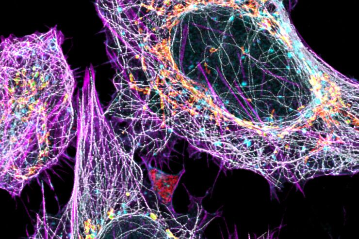
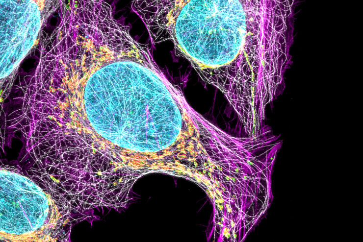
Multicolor Image Gallery
Fluorescence multicolor microscopy, which is one aspect of multiplex imaging, allows for the observation and analysis of multiple elements within the same sample – each tagged with a different…
Loading...

Cancer Research Image Gallery
Fluorescence microscopy allows the study of changes occurring in tissue and cells during cancer development and progression. Techniques such as live cell imaging are critical to understand cancer…
Loading...
Cell Biology Image Gallery
Cell biology studies the structure, function and behavior of cells, including cell metabolism, cell cycle, and cell signaling. Fluorescence microscopes are an integral part of a cell biologist…
Loading...

Developmental Biology Image Gallery
Developmental biology explores the development of complex organisms from the embryo to adulthood to understand in detail the origins of disease. This category of the gallery shows images about…
Loading...

From Light to Mind: Sensors and Measuring Techniques in Confocal Microscopy
This article outlines the most important sensors used in confocal microscopy. By confocal microscopy, we mean "True Confocal Scanning", i.e. the technique that illuminates and measures one single…
