
Medical Specialties
Medical Specialties
Explore a comprehensive collection of scientific and clinical resources tailored for HCPs, including peer insights, clinical case studies, and symposia. Designed for neurosurgeons, ophthalmologists, and specialists in Plastic and Reconstructive surgery, ENT, and dentistry. This collection highlights the latest advancements in surgical microscopy. Discover how cutting-edge surgical technologies, such as AR fluorescence, 3D visualization, and intraoperative OCT imaging, empower confident decision-making and precision in complex surgeries.
Filter articles
Tags
Products
Loading...

Efficient Long-term Time-lapse Microscopy
When doing time-lapse microscopy experiments with spheroids, there are certain challenges which can arise. As the experiments can last for several days, prolonged sample survival must be achieved…
Loading...
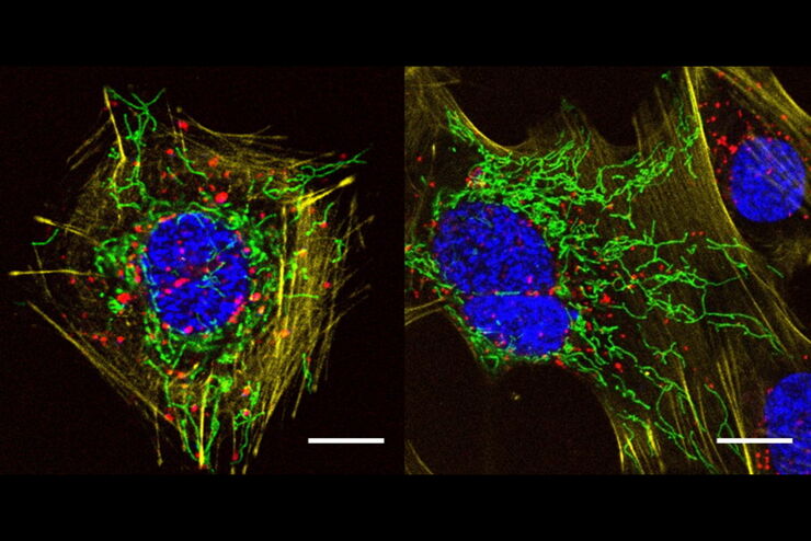
A Versatile Palette of Fluorescent Probes
Researchers at the Max Planck Institute for Medical Research in Heidelberg have developed a general strategy to synthesize live-cell compatible fluorogenic probes, and the result are the new MaP (Max…
Loading...
 and mito OM (red) in a live U2OS cell")
Multicolor 4D Super Resolution Light Sheet Microscopy
The AI Microscopy Symposium offers a unique forum for discussing the latest AI-based technologies and tools in the field of microscopy and biomedical imaging. In this scientific presentation, Yuxuan…
Loading...

Hyperplex Cancer Tissue Analysis at Single Cell Level with Cell DIVE
The ability to study how lymphoma cell heterogeneity is influenced by the cells’ response to their microenvironment, especially at the mutational, transcriptomic, and protein levels. Protein…
Loading...

How to Prepare your Specimen for Immunofluorescence Microscopy
Immunofluorescence (IF) is a powerful method for visualizing intracellular processes, conditions and structures. IF preparations can be analyzed by various microscopy techniques (e.g. CLSM,…
Loading...

Live-Cell Imaging Techniques
The understanding of complex and/or fast cellular dynamics is an important step for exploring biological processes. Therefore, today’s life science research is increasingly focused on dynamic…
Loading...

Fluorescent Dyes
A basic principle in fluorescence microscopy is the highly specific visualization of cellular components with the help of a fluorescent agent. This can be a fluorescent protein – for example GFP –…
Loading...
The AI-Powered Pixel Classifier
Achieving reproducible results manually requires expertise and is tedious work. But now there is a way to overcome these challenges by speeding up this analysis to extract the real value of the image…
Loading...

Using Machine Learning in Microscopy Image Analysis
Recent exciting advances in microscopy technologies have led to exponential growth in quality and quantity of image data captured in biomedical research. However, analyzing large and increasingly…
